
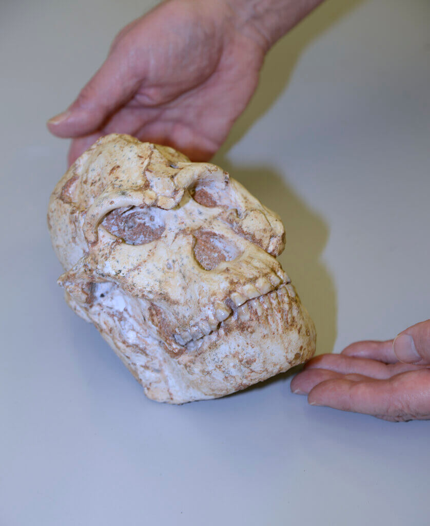
OXFORDSHIRE, England — A near-complete fossil skeleton of an early human found in a South African cave during the 1990s was famously given the name “Little Foot” after its discovery. To say Little Foot has been a hugely important puzzle piece in human is an understatement. Now, scientists are hailing the results of a high-tech scan of the Australopithecus skeleton, which reveal more insight into our species’ origins.
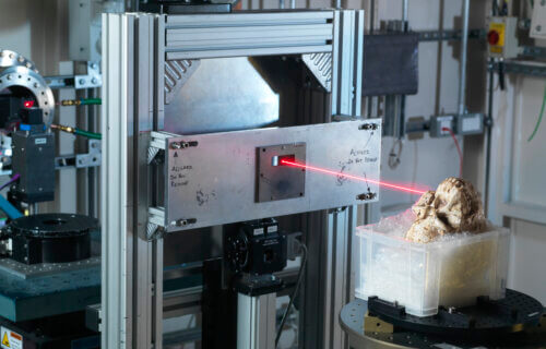
In 2019, a research team brought Little Foot’s skull to Diamond Light Source in the United Kingdom, one of the most advanced scientific facilities in the world. Images of the 3.67-million-year-old fossil revealed details and high-resolution analysis of the bony structures. The researchers say this skull is a great candidate for learning about human origins and evolution since it is amazingly complete.
“We had the unique opportunity to look at the finest details of the craniodental anatomy of the Little Foot skull. While scanning it, we did not know how well the smallest structures would be preserved in this individual, who lived more than 3.5 million years ago. So, when we were finally able to examine the images, we were all very excited and moved to see such intimate details of the life of Little Foot for the first time,” says lead author and principal investigator, Dr. Amelie Beaudet, from the University of Cambridge, in a statement.
“The microstructures observed in the enamel indicate that Little Foot suffered through two clear periods of dietary stress or illness when she was a child,” adds Dr. Beaudet. She and her team were also able to see detailed images of the microscopic canals in the jaw bone that once housed blood vessels.
According to Dr. Beaudet, the structure of these canals indicate a period of remodeling suggesting a change in diet as well as the age at which Little Foot died.
The observations also included channels that were less than 1 mm wide thought to have helped to regulate the temperature of the brain. The team explained that since evolution has increased the size of human brains, these findings could help in understanding the evolution of thermoregulation in the brain as well. “Traditionally, none of these observations would have been possible without cutting the fossil into very thin slices, but with the application of synchrotron technology there is an exciting new field of virtual histology being developed to explore the fossils of our distant ancestors,” said Dr. Beaudet.
“Important aspects of early hominin biology remain debated, or simply unknown. In that context, synchrotron X-ray imaging techniques like microtomography have the potential to non-destructively reveal crucial details on the development, physiology, biomechanics, and taxonomy of fossil specimens,” explains Dr. Thomas Connolley, principal beamline scientist at Diamond. “Little Foot’s skull was also scanned using the adjacent IMAT neutron instrument at ISIS Neutron and Muon Source, combining X-ray and neutron imaging techniques in one visit to the UK. With such a rich volume of information collected, we’re eager to make more discoveries in the complementary X-ray and neutron tomography scans.”
“This level of resolution is providing us with remarkably clear evidence of this individual’s life. We think there will also be a hugely significant evolutionary aspect, as studying this fossil in this much detail will help us understand which species she evolved from and how she differs from others found at a similar time in Africa. Funding permitting, we hope to be able to bring other parts of Little Foot to Diamond,” says principal investigator, and associate professor, Dominic Stratford, from the University of Witwatersrand School of Geography, Archaeology and Environmental Studies.
“This research was about bringing the best-preserved Australopithecus skull to the best of the best synchrotron facility for our purposes. Traditionally, hominins have been analyzed by measuring and describing the exterior shapes of their fossilized bones to assess how these differ between species. Synchrotron development and microCT resources mean that we are now able to virtually observe structures inside the fossils, which hold a wealth of information,” says Dr. Stratford.
“It has taken us 23 years to get to this point. This is an exciting new chapter in Little Foot’s history, and this is only the first paper resulting from her first trip out of Africa. We are constantly uncovering new information from the wealth of new data that was obtained,” adds Professor Ron Clarke, the British scientist based in South Africa who discovered and excavated Little Foot.
This study is published in eLife.