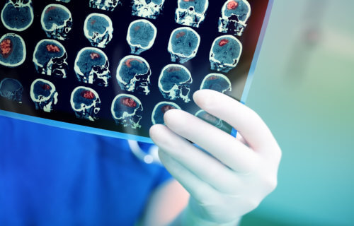
PHILADELPHIA, Pa. — Serious head trauma seldom ends with the injury itself. Patients often experience long-lasting effects that disrupt their lives. Groundbreaking new research finds MRI brain scans after a head injury may prove to be the long-sought key to identifying post traumatic stress disorder (PTSD) early. The study reveals that a specific marker in the brain has a connection to the condition.
PTSD is still very much a puzzle to modern scientists and doctors. While most people probably associate PTSD with returning veterans, this awful psychiatric disorder can occur in anyone exposed to a severe physical or psychological event. Common symptoms include depression, cognitive disturbances, and extreme anxiety.
However, not everyone who encounters something awful will develop PTSD. This has represented a major medical roadblock for years. The ability to accurately predict the onset of PTSD would open up a world of improved treatment options and a better chance at overcoming the condition. Now, it appears MRI scans following a traumatic brain injury (TBI) are the answer to this problem. Researchers report such scans can reveal potential brain biomarkers of PTSD.
“The relationship between TBI and PTSD has garnered increased attention in recent years as studies have shown considerable overlap in risk factors and symptoms,” says lead author Murray Stein, MD, MPH, FRCPC, a Distinguished Professor of Psychiatry and Family Medicine & Public Health at the University of California San Diego, in a media release. “In this study, we were able to use data from TRACK-TBI, a large longitudinal study of patients who present in the Emergency Department with TBIs serious enough to warrant CT (computed tomography) scans.”
Predicting when PTSD will develop
Study authors examined over 400 TBI patients during this study. Along with an performing MRIs, they tested each person for PTSD three and six months after their injuries. At the three month mark, 77 patients (18%) showed signs of likely PTSD. After six months, 70 patients still showed the same indications (16%).
“MRI studies conducted within two weeks of injury were used to measure volumes of key structures in the brain thought to be involved in PTSD,” Dr. Stein explains. “We found that the volume of several of these structures were predictive of PTSD 3-months post-injury.”
Digging into these findings in greater detail, at the three month mark smaller than usual brain volume within the cingulate cortex, the superior frontal cortex, and the insula all predicted PTSD. Those brain regions help regulate arousal, attention, and emotional control.
Notably, MRI scans did not predict PTSD at the six month mark.
The first step toward a new treatment
These aren’t the first findings to observe a connection between lower brain volume and PTSD. Prior research has concluded reduced cortical volume is likely a major PTSD risk factor. Researchers theorize a “brain reserve” (higher cortical volumes) can provide a certain degree of extra protection or resilience against the condition.
As of now, these findings aren’t definitive enough to start being applied in hospitals all over. Study authors are confident, however, that their work is the first step toward a better understanding of PTSD and how to quickly identify and treat the disorder.
“It does pave the way for future studies to look even more closely at how these brain regions may contribute to (or protect against) mental health problems such as PTSD,” Dr. Stein adds.
“This very important study uses magnetic resonance imaging to take the field a step closer to understanding why some people develop PTSD after trauma and others do not. It also lays the groundwork for future research aimed at using brain imaging to help predict that a person is at increased risk and may benefit from targeted interventions to reduce the clinical impact of a traumatic event,” concludes Dr. Cameron Carter, the editor of this study’s publishing journal.
The study is published in Biological Psychiatry: Cognitive Neuroscience and Neuroimaging.