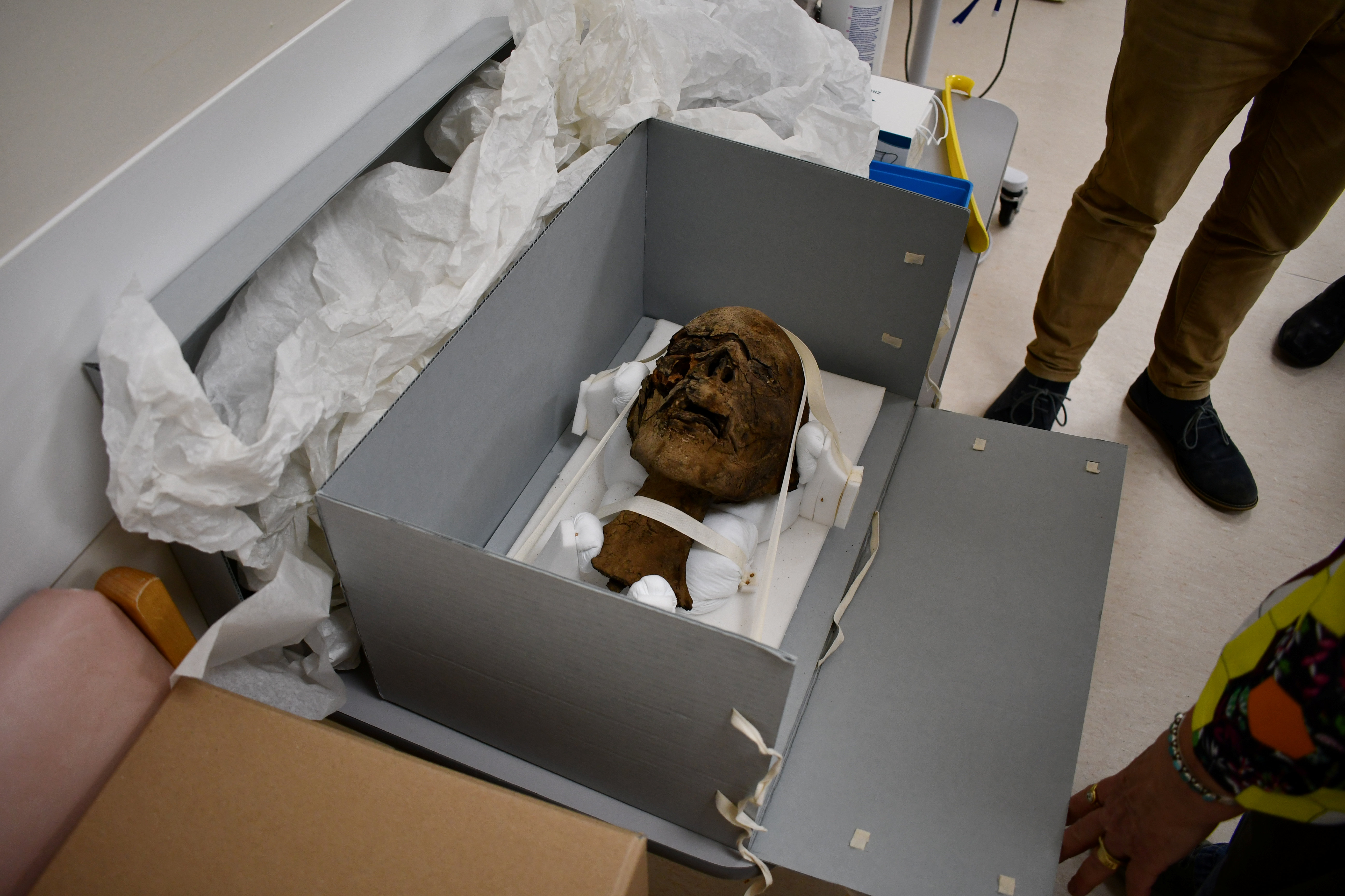

CANTERBURY, United Kingdom — A decapitated Egyptian mummy’s head has been discovered in an attic in the British county of Kent. Now, scientists are putting it through a state-of-the-art CT scan to try and uncover its secrets.
The head, which is at least 2,000 years-old, may have been a “souvenir,” brought to England in the 19th century and given to the home’s owner in the early 20th century. It was discovered in a glass case during a clear-out after the death of a man in Ramsgate.
Researchers say the head, minus its body and any wrappings, was inherited by the man’s brother, who took it to the Canterbury Museums and Galleries for examination.
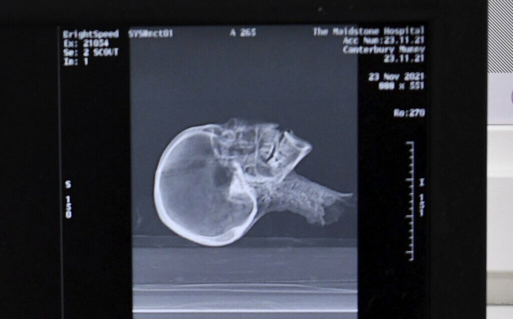
Initial X-rays at Canterbury Christ Church University suggested that it was an adult woman, however, the team organized a detailed CT scan to learn more about the long-dead individual.
Preliminary results from the scan, carried out at Maidstone Hospital, indicate that the brain has been removed, the teeth are worn down by a rough diet, and the tongue actually shows remarkable preservation.
There appears to be tubing of unknown material within the left nostril and in the spinal canal. Whether this something ancient from the mummification process or something more recent origin is unknown.
Could scientists piece this person’s face back together?
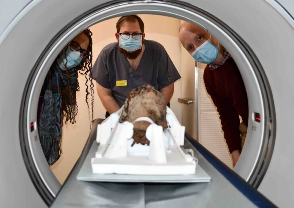
The team is hoping to eventually recreate the face of the mysterious mummy and reconstruct the hidden history of this Egyptian.
“The scan provides a huge amount of information – everything from dental status, pathologies, method of preservation as well as assisting estimations of age and sex. We plan on using the scanning data to create a three-dimensional replica of the head and possible facial reconstruction to allow a more intensive study of it without exposing the actual artifact,” says James Elliott, a senior radiographer at Maidstone and Tunbridge Wells NHS Trust, in a media release.
“The head was found in the attic of a house in Kent which was being cleared out following the death of the owner. During the Victorian times items like this used to be brought back from Egypt as souvenirs and may well have been passed down through generations to the person who owned it.”
Since the head was donated without any details about how it came into its late owner’s possession, very little is actually known about the head. A CT scanner typically helps to identify and diagnose various conditions, including broken bones, internal organ injuries, and signs of stroke. It also can help determine the location, size, and shape of a cancerous tumor before radiotherapy.
CT scans are reconstructing ancient history
Elliott says mummification was a “common practice” in ancient Egypt, but with the advancement of CT technology, scientists are now able to gather more details about ancient Egyptian traditions.
“Beginning in 3500 BC, mummification was a way to safeguard the spirit in its journey to the afterlife. Mummification was common practice within ancient Egypt for both commoners and royalty, although with different levels of complexity and accompanying wealth,” the researcher explains.
“Ironically, the ancient Egyptians believed that a person’s mind was held in their heart and had little regard for the brain. Regardless of this, the brain was removed to help preservation of the individual. Although traditional accounts state the brain was removed exclusively through the nose, research using CT scans has shown great variability. Until relatively recently, the historic accounts have been accepted as gospel but the scanning of Egyptian mummies has challenged these ideas.”
“This project is part of a larger aim to preserve the head and allow it to be displayed in conservation grade packaging for public viewing,” says Craig Bowen, collections and learning manager at Canterbury Museums and Galleries.
“The conservation process also allows volunteers to experience and take part in important discussions surrounding the preservation, recording and study of human remains.”
Where will the head go now?
The head is being preserved by professional archaeological conservator Dana Goodburn-Brown, ACR, who is also coordinating the research efforts. As part of a collaborative scientific investigation of the head, experts from Canterbury Christ Church University, University of Kent and University of Oxford, will attempt to reconstruct the history of the individual.
The group hopes to present the findings to the public at the Beaney Museum in Canterbury sometime in the near future.
“I feel privileged to be providing care, analysis and liaising with specialists in the multidisciplinary team. Through our sensitively conducted research, we hope to gain a better understanding of the individual to whom the head belonged and to provide appropriate conservation care for their remains,” Goodburn-Brown says.
“In keeping with encouraging Science, Technology, Engineering and Mathematics (STEM), the Beaney Museum will use the application of modern technology with history to enhance learning, widen appeal and increase school visits and outreach.”
“It’s very exciting that Maidstone and Turnbridge Wells NHS Trust is involved with this project. It’s great to see how modern technology can help bring ancient history to life,” concludes Ritchie Chalmers, chief of service for core clinical services. “I, along with the rest of the Trust, look forward to finding out what the CT scan unveils.”
The CT data from this individual will help to understand the wider picture of mummification and will be shared with the IMPACT Mummy Database hosted by Western University.
South West News Service writer Cara Simmonds contributed to this report.
One would assume that it being described as a head it would be decapitated.