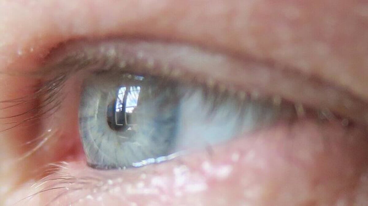
“The eye is our only window into the body, and it’s immune-privileged,” says Anna Herland, senior lecturer in the Division of Bionanotechnology at SciLifeLab at KTH Royal Institute of Technology, and the AIMES research center at KTH and Karolinska Institute. (CREDIT: David Callahan/KTH Royal Institute of Technology)
STOCKHOLM, Sweden — A potential new diabetes treatment may have a certain appeal to a patient’s eyes. Swedish scientists have developed a microscale device that can be implanted in the eye for the treatment of diabetes and other diseases that could benefit from cell-based therapies.
The innovative 3D-printed device, capable of encapsulating insulin-producing pancreatic cells and electronic sensors, was engineered by a team from KTH Royal Institute of Technology and Karolinska Institutet. This collaboration allows for the precise placement of mini-organs, such as pancreatic islets or islets of Langerhans, inside the eye without the need for sutures. This advancement offers exciting prospects for cell-based treatments, particularly for addressing Type 1 or Type 2 diabetes, using the eye as a platform.
Anna Herland, senior lecturer in the Division of Bionanotechnology at SciLifeLab at KTH and the AIMES research center at KTH and Karolinska Institutet, explains that the eye is an ideal location for this technology because it lacks immune cells that could react negatively during the initial stages of implantation. Its transparency allows for visual and microscopic observation of the implant’s behavior over time.
“The eye is our only window into the body, and it’s immune-privileged,” says Herland in a media release.
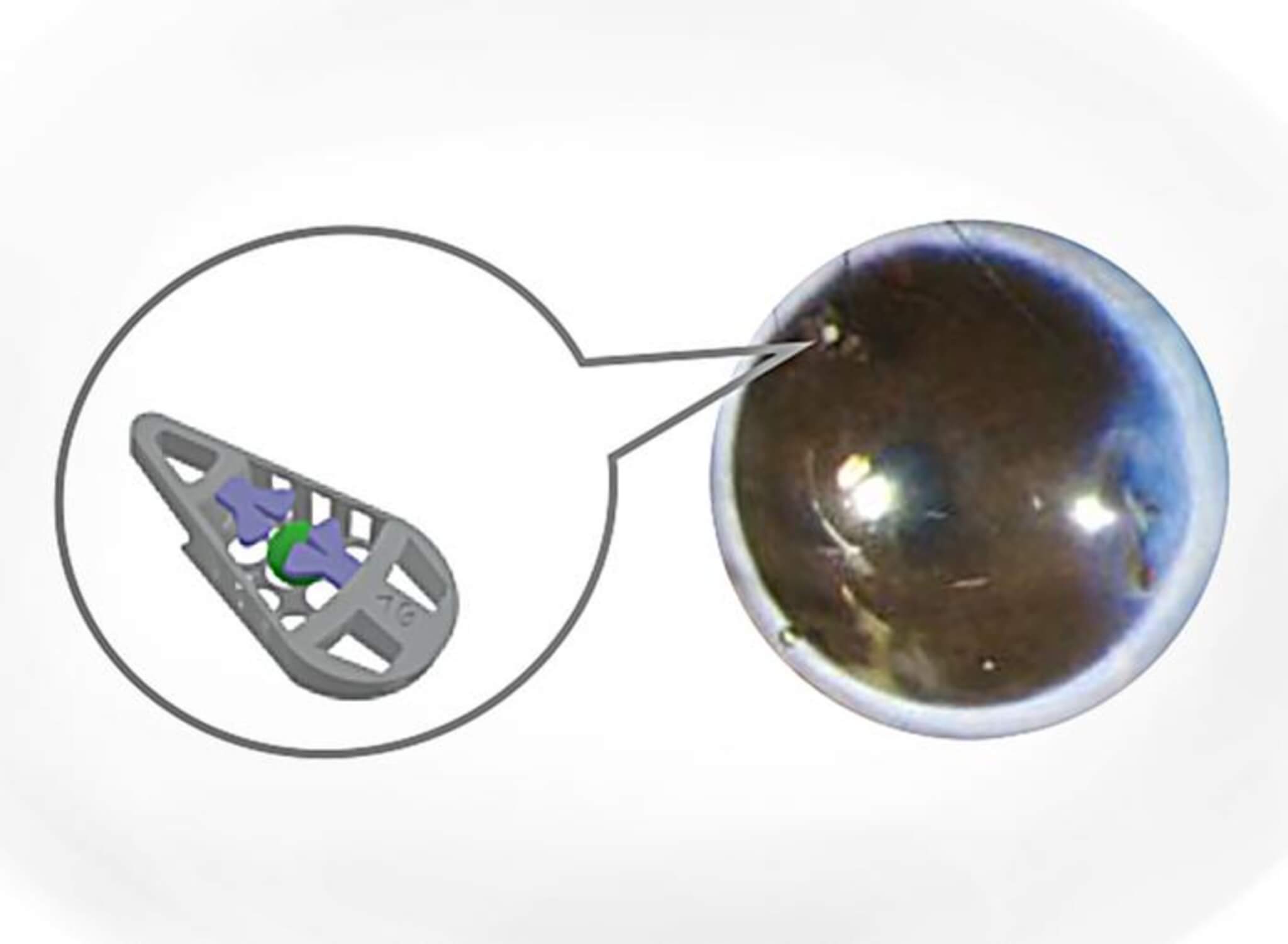
The device itself is designed in the shape of a wedge, measuring around 240 micrometers in length. This design allows the structure to be securely positioned in the anterior chamber of the eye (ACE), at the angle between the iris and the cornea. This work marks the first-ever mechanical fixation of a device in the anterior chamber of the eye.
“We designed the medical device to hold living mini-organs in a micro-cage and introduced the use of a flap door technique to avoid the need for additional fixation,” notes Wouter van der Wijngaart, professor in the Division of Micro- and Nanosystems at KTH.
In tests conducted on mice, the microscale device demonstrated remarkable stability within the living organisms for several months. The mini-organs within the device quickly integrated with the host animal’s blood vessels and functioned as expected.
“The current unit is unique and will among other things form the basis for our continued work to develop an integrated microsystem for studying the function and survival of the islets of Langerhans in the anterior chamber of the eye,” explains Per-Olof Berggren, a professor of experimental endocrinology at Karolinska Institutet. “This is also of great translational importance, as transplantation of Langerhans islands to the anterior chamber of the eye in humans is subject to clinical trials in patients with diabetes.”
Herland highlights that this technology addresses a critical obstacle in the development of cell therapies, including those for diabetes: the need for invasive methods to monitor the graft’s function and guide care to ensure long-term transplant success.
“Ours is a first step towards advanced medical microdevices that can both localize and monitor the function of cell grafts,” says Herland.
The device’s design ensures that it can position mini-organs, such as organoids and islets of Langerhans, without restricting the supply of nutrients to the cells.
“Our design will enable future integration and use of more advanced device functions such as integrated electronics or drug release,” adds Herland.
The study is published in the journal Advanced Materials.
You might also be interested in:
- Implantable ‘oxygen factory’ the size of a coin could replace diabetes insulin injections
- Ozempic and Wegovy protect against Type 2 diabetes, obesity for 3 years
- Best Glucometers: Top 5 Blood Sugar Monitors Most Recommended By Experts
Lea la versión en español en EstudioRevela.com: Pequeño implante ocular podría revolucionar el tratamiento de la diabetes.


