(© unlimit3d - stock.adobe.com)
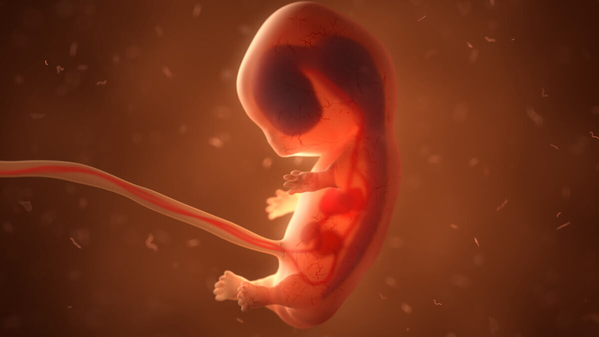
DALLAS — Doctors have successfully completed a groundbreaking brain surgery on a baby in-utero, the first-ever procedure of its kind. The unprecedented surgery fixed potentially deadly damage to blood vessels, saving the infant from suffering heart failure and stroke after birth. The rare prenatal condition is known as Vein of Galen malformation (VOGM), which occurs when arteries carrying high-pressure blood connect to one of the main veins deep at the base of the brain. In normal fetal development, these arteries should link to smaller capillaries, slowing blood flow and delivering oxygen to surrounding tissue.
The U.S. team performed the successful procedure at 34 weeks and two days gestational age using ultrasound-guided transuterine embolization. Due to the rupture of membranes, doctors induced labor, allowing the mother to give birth two days later. The unnamed child is now at home.
“In our ongoing clinical trial, we are using ultrasound-guided transuterine embolization to address the vein of Galen malformation before birth, and in our first treated case, we were thrilled to see that the aggressive decline usually seen after birth simply did not appear,” says lead study author Darren B. Orbach, M.D., Ph.D., co-director of the Cerebrovascular Surgery & Interventions Center at Boston Children’s Hospital and an associate professor of radiology at Harvard Medical School.
Repeated echocardiograms after birth showed marked improvement in cardiac output, and scans displayed normal heart and brain function. Six weeks later, the infant is progressing remarkably well, with no medications, normal eating habits, and weight gain. There are no signs of any negative effects on the brain. The premature newborn did not require any cardiovascular support or surgery following the treatment and spent several weeks in the neonatal intensive care unit. During that time, the newborn had a normal neurological exam and showed no signs of stroke, fluid buildup, or hemorrhage on brain MRIs.
Scroll down to see 5 alternatives to prenatal surgery
Experts described the procedure, detailed in the journal Stroke, as “pioneering.” Professor Gary Satou, a fetal cardiologist at UCLA, says the intervention may be “very impactful” in a specific group of patients.
“As always, a number of these fetal cases will need to be performed and followed in order to establish a clear pattern of improvement in both neurologic and cardiovascular outcomes. Thus, the national clinical trial will be crucial in order to achieve adequate data and, hopefully, successful outcomes,” Satou adds in a media release.
“This is pioneering work being done in a very careful and responsible way,” says Prof. Colin Derdeyn, a neuro-interventional radiologist at the University of Iowa.
However, Derdeyn noted that one successful case is not enough experience to conclude that the risks of this procedure are worth the benefits. Despite that, he adds that the positive hemodynamic changes observed in utero and after birth, such as the reduction in flow, reduction in size of the draining vein, and reversal of the abnormal reversed flow in the aorta, were “really encouraging.”
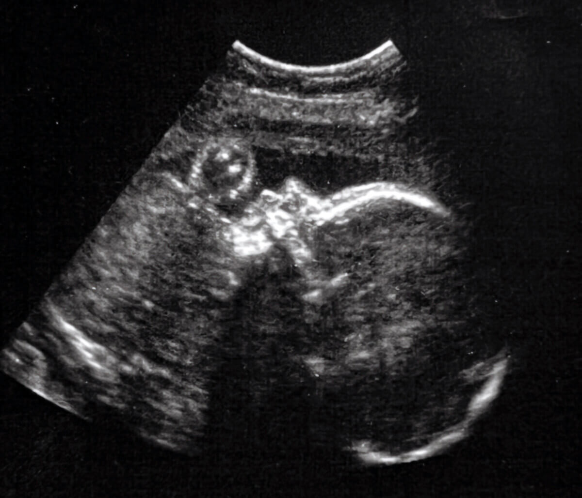
How do doctors find VOGM?
VOGM is most often first seen on a prenatal ultrasound scan and then diagnosed by MRI during the late second or third trimester of pregnancy. It is estimated that VOGM, the most common congenital vascular brain malformation, occurs in as many as one in every 60,000 births.
The current standard of care is treatment after birth with embolization. However, embolization itself is high risk and is not always successful at reversing heart failure. Furthermore, severe brain damage may have already occurred, which may lead to life-long cognitive disabilities and life-threatening conditions for the infant, or even to death.
This innovative approach has the potential to mark a paradigm shift in managing Vein of Galen malformation by repairing the malformation prior to birth and preventing heart failure before it occurs, rather than trying to reverse it after birth.
“This may markedly reduce the risk of long-term brain damage, disability, or death among these infants,” Dr. Orbach says.
The high flow in the malformation has an even more serious effect on the heart and brain after birth, putting enormous pressure on the newborn’s heart and lungs. This may lead to pulmonary hypertension, heart failure, or other life-threatening conditions.
As the national clinical trial progresses, researchers and doctors will continue to gather valuable data on the long-term neurological and cardiovascular outcomes of this groundbreaking intervention. The future success of this procedure could pave the way for new approaches to treating congenital vascular brain malformations and other life-threatening prenatal conditions, ultimately saving lives and improving the quality of life for affected infants and their families.
What are the alternatives to prenatal surgery?
There are alternatives to prenatal surgery for various conditions, including Vein of Galen malformation (VOGM). These alternatives typically involve postnatal treatment and management strategies. Some of the alternatives to prenatal surgery for VOGM and other conditions are:
- Postnatal embolization: For VOGM, the current standard of care involves treating the condition after birth with embolization. This is a catheter-based procedure that closes off the direct artery-to-vein connections, blocking excess blood flow to the brain and heart. However, embolization carries its own risks and may not always be successful in reversing heart failure.
- Surgery: In some cases, surgery may be performed after birth to address specific congenital anomalies. The type of surgery and the approach used will depend on the nature and severity of the condition. For example, surgeries for congenital heart defects may involve repairing or replacing damaged heart structures or creating alternative routes for blood flow.
- Medication: In certain situations, medications may be used to manage symptoms and improve the overall health of the affected infant. For example, drugs to control pulmonary hypertension or heart failure may be prescribed.
- Supportive care: For some conditions, the primary focus may be on providing supportive care to manage symptoms and optimize the infant’s overall health. This can include specialized care in a neonatal intensive care unit (NICU), where infants receive round-the-clock monitoring, respiratory support, and nutritional support as needed.
- Monitoring and follow-up: In less severe cases, doctors may opt to closely monitor the infant’s condition and development, with the possibility of intervening later if the condition worsens or if complications arise.
It is essential to note that the choice of treatment will depend on the specific condition, its severity, and the individual needs of the infant and their family.
South West News Service writer Mark Waghorn contributed to this report.


I really wish that they would not have used a stock photo of a very young (certainly not 36 weeks!) fetus. Perpetuates the false narrative that a baby is just a ‘clump of cells.’ Otherwise this is amazing and wonderful.
This is great news.