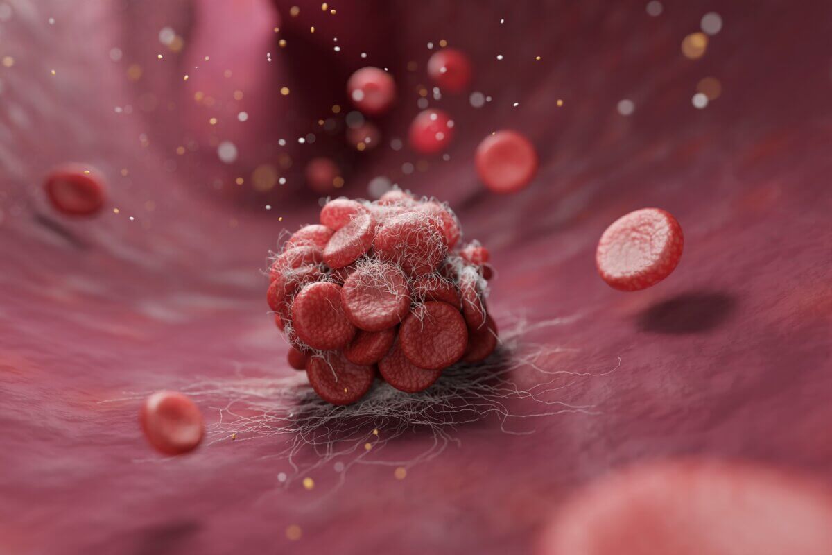
(Image credit: Piyaset on Shutterstock)
Simple red light exposure could slash risk by 75%, mouse model reveals
PITTSBURGH — In hospital rooms across the world, fluorescent lights hum overhead as patients lie in their beds, many at risk for potentially fatal blood clots. But what if those same lights could be modified to help protect patients? A team of researchers has discovered that simply changing the wavelength of light exposure might reduce the risk of dangerous blood clots — and the implications could revolutionize how we think about preventing thrombosis.
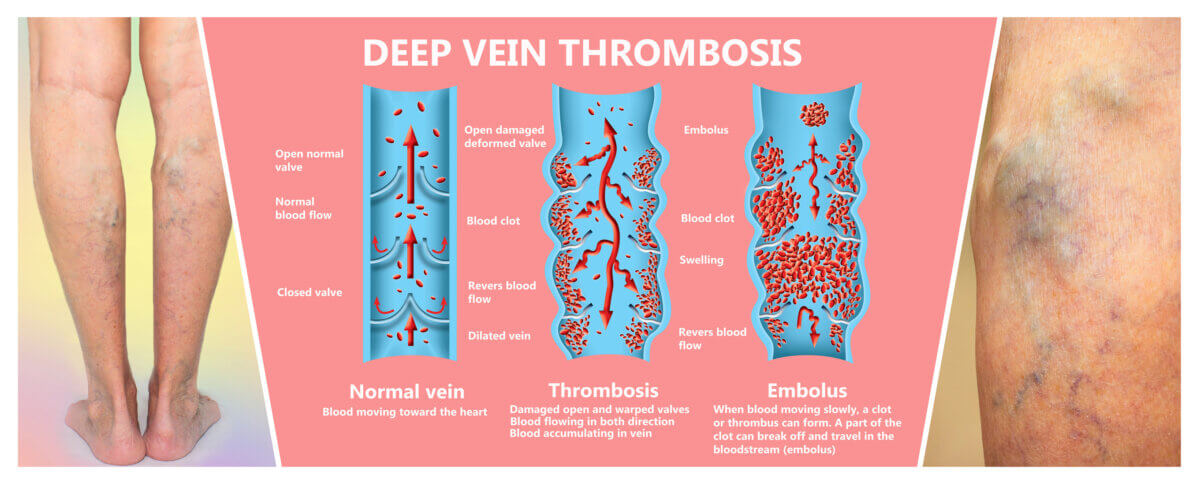
Blood clots affect millions worldwide each year, developing most commonly during long periods of immobility like hospital stays or lengthy flights. Venous thromboembolism (VTE), blood clots that typically form in the deep veins of the legs, strikes up to 10 million people annually and remains the leading cause of preventable hospital death worldwide. Meanwhile, arterial thrombosis, which can lead to strokes and heart attacks, affected over 12 million people globally in 2019 alone.
Current treatments primarily rely on blood thinners, which while effective, carry their own risks and can’t always be used due to bleeding concerns. This limitation has scientists searching for alternative approaches, and new research suggests that something as simple as modifying environmental light exposure could potentially provide an additional tool for preventing dangerous clots.
Scientists have long connected light exposure to health outcomes. The rising and setting of the sun influences metabolism, hormone secretion, and even blood flow. Research has shown that heart attacks and strokes occur more frequently in morning hours than at night. This connection between light and cardiovascular events led Andraska and her colleagues to investigate whether specific types of light could influence blood clot formation.
Led by Dr. Elizabeth A. Andraska from the University of Pittsburgh, researchers from a number of institutions exposed laboratory mice to three different lighting conditions: ambient white light (similar to standard fluorescent lighting), blue light (442 nm wavelength), or red light (617 nm wavelength). After 72 hours of exposure, they examined how the different types of light affected platelets — tiny blood cells that help form clots to stop bleeding — and measured various markers of blood clot formation.
“The light we’re exposed to can change our biological processes and change our health,” says Dr. Andraska, assistant professor of surgery in Pitt’s Trauma and Transfusion Medicine Research Center and vascular surgery resident at UPMC, in a statement. “Our findings could lead to a relatively inexpensive therapy that would benefit millions of people.”
Results, published in the Journal of Thrombosis and Haemostasis, showed mice exposed to red light developed significantly smaller blood clots compared to those exposed to white or blue light. When researchers induced blood clots in the mice’s veins, the clots that formed in red light-exposed mice weighed approximately 4.21 mg compared to 18.54 mg in mice exposed to white light — a reduction of more than 75%. Importantly, the researchers found that activity, sleep, eating, weight and body temperature remained the same between all groups.
The research team observed specific biological changes that help explain these results. Red light exposure was associated with less inflammation and immune system activation. The team found that red light-exposed mice had fewer neutrophil extracellular traps (NETs), web-like structures released by immune cells that normally trap harmful microorganisms but can also contribute to unwanted clot formation. Additionally, mice exposed to red light showed increased fatty acid production, which naturally reduces platelet activation and subsequent clot formation.
Intriguingly, these effects required functioning eyes to work. When researchers repeated their experiments using blind mice, the protective effects of red light disappeared, suggesting that the benefits come from how the body processes light through the visual system rather than from direct effects of light on blood.
Moving from mice to humans, the research team analyzed data from 10,464 cataract surgery patients who received either conventional intraocular lenses or special lenses that filter out approximately 50% of short-wavelength blue light. While the overall results showed no significant difference in blood clot risk between the two groups, cancer patients who received blue light-filtering lenses showed a notably lower risk of developing blood clots. This finding is particularly significant because cancer patients have nine times the risk of blood clots compared to non-cancer patients.
“These results are unraveling a fascinating mystery about how the light to which we’re exposed on a daily basis influences our body’s response to injury,” explains senior author Dr. Matthew Neal, professor of surgery and co-director of the Trauma and Transfusion Medicine Research Center at Pitt.
Looking ahead, the research team is already preparing for clinical trials. They are developing specialized red light goggles to control the amount of light exposure study participants receive and investigating which patients might benefit most from red light therapy. Understanding how red light triggers changes that lower clotting risk could also help scientists develop better medications or therapies that might be more potent and convenient than continuous red light exposure.
“Getting to the bottom of our discovery has the potential to massively reduce the number of deaths and disabilities caused by blood clots worldwide,” says Neal. While further research is needed to fully understand these effects in humans, this relatively simple intervention could represent a significant advance in preventing life-threatening blood clots.
Paper Summary
Methodology
The researchers used a comprehensive approach combining animal studies with human data analysis. In the animal studies, they exposed mice to different types of light for 72 hours, maintaining normal day-night cycles. They then induced blood clots using established laboratory techniques and measured clot formation, platelet function, and various molecular markers. For the human component, they analyzed medical records from cataract surgery patients to compare outcomes between those who received different types of lens implants.
Results
The study found that red light exposure reduced blood clot size by about 77% compared to white light exposure in mice. Platelet aggregation decreased significantly, and markers of inflammation were reduced. In human data, while overall clot risk wasn’t significantly different between lens types, cancer patients with blue light-filtering lenses showed a 47% lower risk of blood clots.
Limitations
The researchers acknowledge several limitations. Mouse retinas differ from human retinas in their sensitivity to red light. The human data was retrospective, meaning it could contain inherent biases. Additionally, the mouse models used artificial methods to induce clots, which may not perfectly mirror natural clot formation in humans.
Discussion and Takeaways
The research suggests that light exposure could be a novel approach to preventing blood clots, particularly in hospital settings. The findings point to an optical pathway rather than direct effects on blood, opening new avenues for research into how the body’s visual system influences blood clotting.
Funding and Disclosures
The research received extensive funding support, including multiple National Institutes of Health grants, support from the University of Pittsburgh Center for Research Computing, National Center for Research Resources Shared Instrumentation grants, an American Heart Association award, and a Physician-Scientist Institutional Award from the Burroughs Wellcome Fund. One author disclosed serving as Chief Medical Officer for Haima Therapeutics and receiving honoraria from several pharmaceutical companies.
Publication Information
This study was published in the Journal of Thrombosis and Haemostasis in January 2025 (Volume 23, Issue 1, pages 123-138), titled “Alterations in visible light exposure modulate platelet function and regulate thrombus formation.”


