
(© twinsterphoto - stock.adobe.com)
OAK BROOK, Ill. – High-tech scans can identify patients at risk of a heart attack years before anything actually happens, a new study reveals. The method called “radiomics” combines data from CT (computed tomography) scans that reveal signs of disease not visible in the images alone.
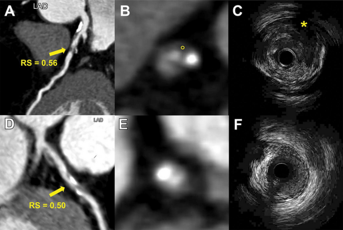
Researchers working with the Radiological Society of North America say the new technique spots deposits of fat in artery walls that could cut off the organ’s blood supply. The large plaques rich in lipids such as bad cholesterol cause most heart attacks. They sometimes rupture but predicting this is challenging.
“The results of this study are encouraging and exciting,” says study co-lead author Long Jiang Zhang, M.D., Ph.D., from the Department of Radiology at Jinling Hospital, Medical School of Nanjing University, in a media release. “Radiomics provided a more accurate approach to detect vulnerable plaques compared to conventional coronary CT angiography anatomical parameters.”
Radiomics is a potential gamechanger, enabling the use of statins or other protective drugs at the earliest opportunity. The Chinese team developed a model based on information from the CT scans of around 300 individuals. It enabled detection of life-threatening clots in just over 700 patients with suspected coronary artery disease.
A “high radiomic signature” displayed an independent link to potential heart attacks over an average follow-up period of three years. It would be easy to add into practice, helping classify those likely to suffer such an event in the future.
“If the radiomics analysis is embedded into the routine CT angiography workstation, it can automatically identify vulnerable plaques for clinician review,” Dr. Zhang says. “Thus, radiomics may significantly improve the accuracy and precision of high-risk plaque detection in routine clinical practice.”
According to the Centers for Disease Control and Prevention, someone in the United States has a heart attack every 40 seconds. Dr. Zhang’s system would enable hundreds of thousands of people over 40 to undergo predictive CT scans — particularly smokers, diabetics, and those who are overweight.
Previous research suggests that one in 10 of those who receive an “all-clear” diagnosis after a regular CT scan are still at risk of a heart attack because of plaques the scan did not detect. Dr. Zhang and the team are now building a radiomics model from different scanner types and vendors. They are also planning a larger study of 10,000 patients.
“With the support of large observational studies and randomized controlled trials, the radiomics approach may help guide clinical decision-making and improve patient care in the future,” the researcher concludes.
The study is published in the journal Radiology.
South West News Service writer Mark Waghorn contributed to this report.


