(Credit: fotovapl/Shutterstock)
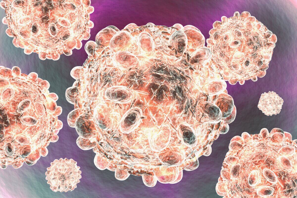
COPENHAGEN, Denmark — When people think of life-threatening viruses like COVID, HIV, and rabies, the only reassurance is that there are some types of treatment available to prevent fatal symptoms or reduce the spread to others. However, this isn’t the case for the roughly 58 million people who deal with the chronic inflammation stemming from hepatitis C.
Now, Danish researchers have taken a step in the right direction toward treating this problem. Scientists have identified the protein complex that gives the hepatitis C virus the ability to bind and infect cells. This bit of cellular knowledge was an ongoing issue in the race to create a hepatitis C vaccine.
“We are the first ever to identify the protein complex at the surface of the hepatitis C virus that enables it to bind to our cells,” says senior study author Jannick Prentø, an associate professor at the University of Copenhagen, in a media release.
No treatment has successfully eradicated hepatitis C from the world. The disease leads to 300,000 deaths yearly. It is also one of the leading causes of liver cancer.
Published in the journal Nature, researchers outlined how two envelope proteins combine. The molecular arrangement of these proteins allows them to infect cells and evade detection from neutralizing antibodies. Though the protein structure is different, it acts similarly to the coronavirus spike protein in penetrating and hijacking control of human cells.
“This knowledge of the structure of the protein complex will enable us to design vaccine candidates that can prevent the virus from infecting the cells,” adds Elias Augestad, a postdoc at the University of Copenhagen and lead study author.
Understanding the protein complex and how the hepatitis C virus infects cells is the last crucial step in designing a vaccine. The proposed vaccine would ideally train the immune system to identify the hepatitis C protein complex, prompting the release of neutralizing antibodies. These antibodies would then bind to the protein complex, making it unable to bind to cells.
“Expressing and cleaning up the protein complex is extremely difficult, which is why it has not been done before. The structure of these proteins on the surface of the hepatitis C virus makes them extremely vulnerable,” Prentø explains. “Researchers did not know what they were dealing with, and therefore, whenever someone tried to reproduce these protein structures in the lab they would fall apart before they could get a chance to study them.”
According to the authors, replicating and studying the protein complexes in hepatitis C viruses is necessary for vaccine development. With scientists now having a better knowledge of the structure that makes the hepatitis C virus so infectious, the hope for a new medical breakthrough is closer than ever.
Paper Summary
Methodology
The researchers studied the structure of proteins on the surface of the hepatitis C virus (HCV). They used a special microscope called a cryo-electron microscope to take very detailed pictures of these proteins. To do this, they first had to make and purify the proteins in a lab. They then froze the proteins very quickly and took many pictures of them. Using computers, they combined these pictures to create a 3D model of the proteins.
Key Results
The study found that the surface proteins of HCV, called E1 and E2, form pairs that then join together in twos. This structure helps explain how the virus avoids detection by our immune system. The researchers also discovered how certain parts of these proteins, which were previously hard to see, are arranged. This includes a part that might help the virus enter our cells. Understanding this structure could help scientists design better vaccines against HCV.
Study Limitations
The proteins were studied outside of the virus, which might not perfectly represent how they look on a real virus. The structure was frozen for study, so it doesn’t show how the proteins might move or change shape. The researchers only looked at proteins from two types of HCV, but there are many more types that might be slightly different. Some parts of the proteins were still hard to see clearly, even with this advanced method.
Takeaways & Discussion
This study provides the first detailed look at how HCV surface proteins are arranged in pairs of pairs (homodimers). The structure helps explain how HCV avoids antibodies that could fight the infection. The researchers found a possible “hidden” part of the protein that might be important for the virus to infect cells. This information could be very useful for developing new vaccines against HCV. The structure also suggests that the virus might need to change shape significantly to infect cells, which wasn’t known before.
Funding & Disclosures
The study was funded by several organizations, including the Novo Nordisk Foundation, the Lundbeck Foundation, and the Candys Foundation. The researchers used facilities at the University of Copenhagen and in Stockholm, Sweden. The authors declared that they have no competing interests, which means they don’t have any financial or personal relationships that could have influenced their research.


