
(ID 343128957 © Prostockstudio | Dreamstime.com)
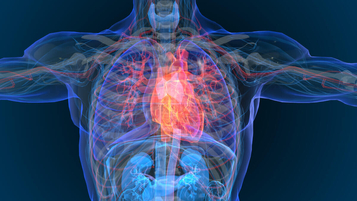
CHARLOTTESVILLE, Va. — Your heart harbors a secret: a layer of fat that can either protect it or potentially contribute to its demise. Now, University of Virginia researchers have invented a revolutionary MRI technique that allows them to study this enigmatic tissue in unprecedented detail, potentially opening new frontiers in cardiac medicine.
This innovative technique examines epicardial adipose tissue (EAT), a specialized fat layer that wraps around our hearts. While EAT naturally serves protective functions in healthy individuals, providing both energy reserves and thermal insulation for the heart, it can undergo concerning changes under certain conditions.
The research team, led by Dr. Frederick H. Epstein, of UVA’s Department of Biomedical Engineering, developed this specialized MRI technique to examine the fatty acid composition of EAT, measuring different types of fats: saturated, monounsaturated, and polyunsaturated fatty acids.
“Using this new MRI technique, we now for the very first time have the ability to know the composition of the fat that accumulates around the heart. This is important because depending on its makeup, the fat which surrounds the heart has the potential to release damaging substances directly into the heart muscle, leading to serious heart problems,” explains researcher Amit R. Patel, MD, a cardiologist and imaging expert at UVA Health.
To test their new imaging method, the researchers studied 21 patients who had experienced ST-segment elevation myocardial infarction (STEMI), a severe type of heart attack. All patients were examined within 96 hours after blood flow was restored to their hearts. The study group included 14 men and seven women, with an average age of 62.6 years.
The technique provides detailed insights into EAT composition that weren’t previously possible with conventional imaging. Understanding the proportions of different fat types is crucial because each affects heart health differently. For instance, saturated fats have been linked to inflammatory responses in adipose tissue, while certain unsaturated fats may help counter inflammation.
One of the major technical achievements was developing a method that could capture clear images despite the constant motion of the heart and lungs. “The ability to make these measurements in epicardial adipose tissue required the use of advanced computational methods that can extract the unique signature of saturated fatty acids from an overall noisy signal,” explains Epstein, who also serves as associate vice president for research at UVA.
The study found distinct differences in EAT composition compared to other fat deposits in the body. Notably, the researchers discovered significant correlations between EAT composition and measures of heart function and structure in STEMI patients. The findings suggest that analyzing EAT composition might provide valuable insights into cardiac health and disease progression.

Looking ahead, Dr. Patel envisions potential therapeutic applications. “With our ongoing research, we hope to show that we can convert the unhealthy fat which surrounds the heart to a more healthy type of fat with either diet and exercise or through the use of medications. We believe that by doing so, we will be able to reduce some of the complications associated with heart disease.”
From protective cushion to potential indicator of heart health, the fat surrounding our hearts may hold crucial clues about cardiac disease. This new imaging technique brings us closer to understanding its complex role.
Paper Summary
Methodology
The researchers developed a specialized MRI sequence that captures multiple images of the heart area during a single breath-hold. The technique uses a complex mathematical model to analyze how different types of fat molecules respond to magnetic fields, allowing them to distinguish between various fatty acids. The images are then processed using advanced computer algorithms to reduce noise and improve clarity, resulting in detailed maps of fat composition.
Results
The study found that EAT had significantly lower levels of polyunsaturated fats compared to other body fat deposits. Importantly, patients with higher levels of saturated fats in their EAT showed larger heart chambers and reduced heart function. The technique proved highly accurate when tested on oil phantoms (test objects), showing strong correlations with known fat compositions.
Limitations
The study focused specifically on STEMI patients examined within a narrow time window after their heart attacks, so findings may not generalize to other populations or cardiac conditions. The sample size was relatively small at 21 patients. The technique requires sophisticated MRI equipment and complex data processing, which might limit its widespread adoption. Additionally, the imaging method works best on larger deposits of fat tissue and may be less accurate for very thin EAT layers.
Discussion and Takeaways
The research suggests that the composition of fat around the heart, not just its quantity, may play a crucial role in heart disease. The ability to analyze this fat non-invasively could lead to better understanding of heart disease progression and potentially new treatment strategies. The technique might also help identify patients at higher risk of heart problems before they develop severe symptoms.
Funding and Disclosures
The study received substantial support from multiple prestigious organizations, including the National Heart, Lung, and Blood Institute (grant R01 HL162872), the National Institute on Aging (grant R01 AG076360), the American Heart Association (grant 23PRE1011202), and the National Institutes of Health (grant T32 HL007284). The researchers disclosed all their funding sources and declared no conflicts of interest. This extensive funding underscores the perceived importance and potential impact of this research in the field of cardiac medicine.


