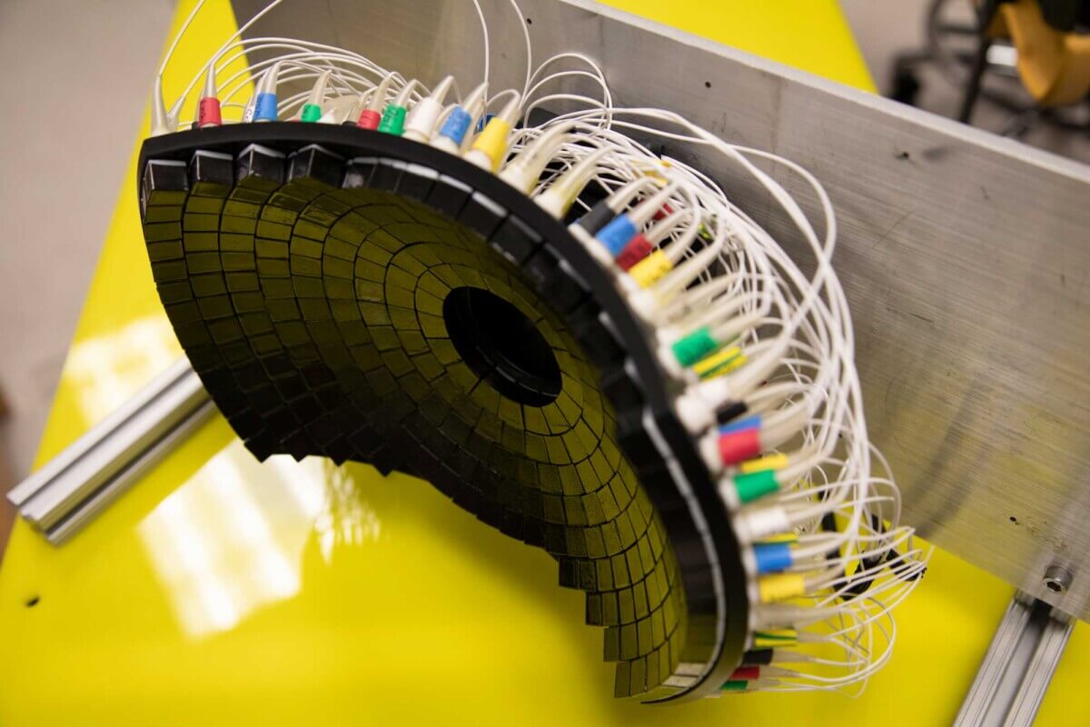
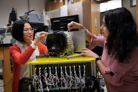
The 700kHz, 260-element histotripsy ultrasound array transducer used in Prof. Xu’s lab. (Image credit: Marcin Szczepanski, Michigan Engineering)
ANN ARBOR, Mich. — Scientists from the University of Michigan have found an inventive way of getting rid of liver tumors for good. Their latest animal study shows that noninvasive sound technology effectively destroys 50 to 75 percent of the cancerous mass. By destroying a majority of the tumor, the immune system is capable of eliminating the rest and preventing the disease from spreading.
Liver cancer is one of the top 10 causes of cancer-related deaths worldwide. Despite multiple treatment options, it continues to have a five-year survival rate of less than 18 percent in the United States. There is also a high rate of tumor recurrence and high risk of it spreading to other parts of the body. However, in the current study, over 80 percent of mice exposed to targeted sound waves did not show any evidence of the tumor coming back.
“Even if we don’t target the entire tumor, we can still cause the tumor to regress and also reduce the risk of future metastasis,” says study co-author Zhen Xu, professor of biomedical engineering at Michigan, in a university release.
How does histotripsy work?
The sound treatment known as histotripsy uses ultrasound waves to physically destroy the targeted tissue with millimeter precision. It also stimulated the rats’ immune system to respond and contribute to the regression of the untargeted portion of the tumor.
“Histotripsy is a promising option that can overcome the limitations of currently available ablation modalities and provide safe and effective noninvasive liver tumor ablation,” says Tejaswi Worlikar, a doctoral student in biomedical engineering. “We hope that our learnings from this study will motivate future preclinical and clinical histotripsy investigations toward the ultimate goal of clinical adoption of histotripsy treatment for liver cancer patients.”
While ultrasounds use sound waves to produce images of the body, the researchers modified sound waves for use as an actual treatment. A benefit to histotripsy in the study was that it did not produce harmful side-effects, which are common with cancer treatments such as radiation and chemotherapy.
“Our transducer, designed and built at U-M, delivers high amplitude microsecond-length ultrasound pulses—acoustic cavitation—to focus on the tumor specifically to break it up,” Dr. Xu explains. “Traditional ultrasound devices use lower amplitude pulses for imaging.”
Histotripsy uses microsecond long pulses to create microbubbles within targeted tissues. The bubbles rapidly expand and collapse. These violent and mechanical stressors destroy enough cancer cells to break up the tumor’s structure.
Scientists are currently testing the new sound technique in a human liver cancer clinical trial in the United States and Europe.
The study is published in the journal Cancers.


Can you sign for clinical trial
Where can sign up now
Can you sign up for clinical trial