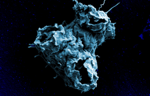
AMHERST, Mass. — To understand how life became what it is today, there may be no better answer than a brain-eating amoeba. Researchers at the University of Massachusetts Amherst are studying an organism called Naegleria and believe it may offer new insight into the evolution of life on Earth.
Scientists investigated how Naegleria evolved to develop microtubules that help the cell complete internal tasks from transporting food and waste to providing structural support. The findings may also help in devising treatment strategies against brain-eating amoebas.
Life relies on microtubules made up of sets of tubulins. Along with maintaining the daily functions of the cell, the microtubules help with dividing and replicating the cell. More specifically, there is a stage in mitosis where microtubules pull chromosomes to opposite sides of the cell, dividing them into two identical sets.
Previously, biologists assumed the brain-eating amoeba used a specific type of tubulin during cell division. But in the new study, the team showed that Naegleria evolved to have three different sets of tubulins. One pair of tubulins only helps during mitosis while the other, called flagellate tubulin, is in charge of cellular movement.
In the study, the team compared the tubulins in Naegleria and the structures they build with those seen in other species. “If we can understand the basic biology of Naegleria,” says Katrina Velle, a postdoctoral scholar of biology at UMass Amherst and the lead author of the study, in a statement. “We can learn how to kill it by devising drugs that target the amoeba’s unique tubulins.”
Naegleria may also give researchers a glimpse into the basic rules that have driven life’s evolutionary progress. “All organisms have to replicate themselves,” says Lillian Fritz-Laylin, a professor of biology at UMass Amherst and a senior author of the paper. “We know how the replication processes works for some cells, but there’s a huge set that we don’t understand. Naegleria lets us test the rules scientists have come up with to see if they hold here.”
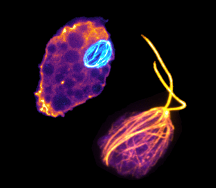
The team used microscopes to study the Naegleria cells they grew in the lab. These cells were stained with different chemicals to identify the tubulins, emitting a glowing color. The microscopes helped them take extremely high-resolution 3-D photographs that allowed them to measure, count, and analyze the different microtubule structures.
“People often think of technology driving science,” adds Fritz-Laylin. “But in this case, the questions we are trying to answer are so fundamental to how life on earth operates, and of such interest to so many scientific specialties, that we needed to assemble an international team of various experts. In this case, collaboration, teamwork and effective communication drove the science.”
The study is published in the journal Current Biology.