SEATTLE — The interior design of human cells has been mapped for the first time in a breakthrough that could revolutionize healthcare.
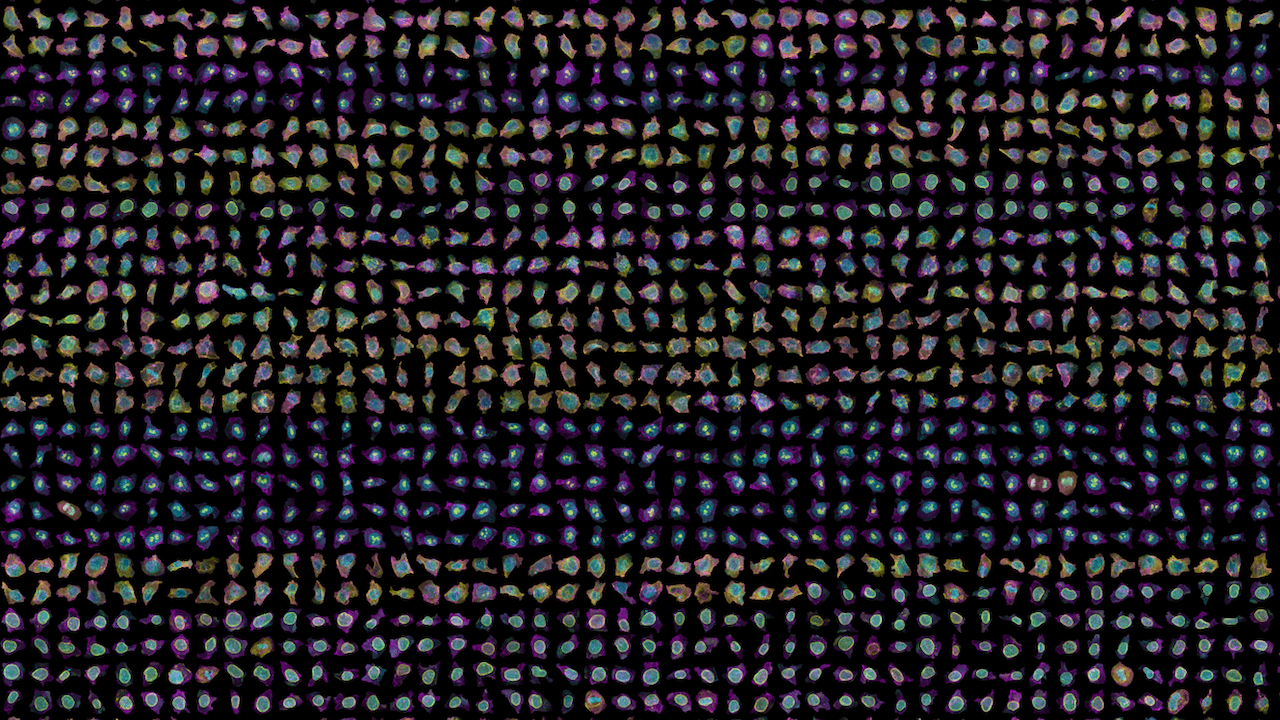
A database of 200,000 images is shedding fresh light on the building blocks of life, according to a team at the Allen Institute for Cell Science. The images reveal details about the rich variation in each cell’s shape, even among genetically identical cells grown under the same conditions.
The breakthrough offers hope of new treatments for cardiovascular disease, cancer, dementia, and other serious illnesses.
“The way cells are organized tells us something about their behavior and identity,” says study leader Susanne Rafelski, Ph.D., Deputy Director of the Allen Institute for Cell Science, in a media release. “What’s been missing from the field, as we all try to understand how cells change in health and disease, is a rigorous way to deal with this kind of organization. We haven’t yet tapped into that information.”
The human body has approximately 37 trillion cells. Working out what they all do will transform medical research. The study, published in Nature, provides a roadmap for biologists, according to Dr. Rafelski. It also reveals some key principles of the cells the team is analyzing.
Cells come in all shapes and sizes
Known as human induced pluripotent stem cells (iPSCs), they are derived from skin or blood cells. Reprogramming them back into an embryonic-like state enables development of an unlimited source of any type of human cell needed for therapeutic purposes.
“Part of what makes cell biology seem intractable is the fact that every cell looks different, even when they are the same type of cell. This study from the Allen Institute shows that this same variability that has long plagued the field is, in fact, an opportunity to study the rules by which a cell is put together,” says Wallace Marshall, Ph.D., a professor of Biochemistry and Biophysics at UC San Francisco and a member of the Allen Institute’s Scientific Advisory Board. “This approach is generalizable to virtually any cell, and I expect that many others will adopt the same methodology.”
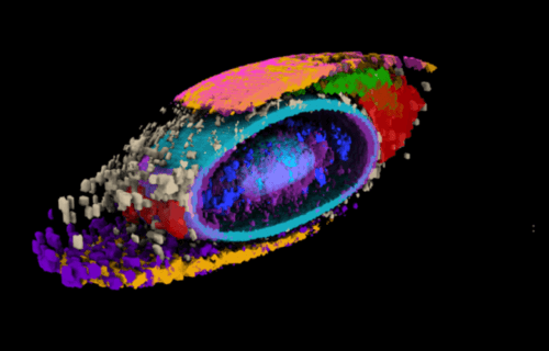
Dr. Rafelski and the team first built a collection of stem cells genetically engineered to light up different internal structures under a fluorescent microscope. With cell lines in hand that label 25 individual structures, the scientists then captured high-resolution, 3D images of more than 200,000 different cells.
Using computational analyses, they developed a “shape space” that objectively describes each stem cell’s eight different external dimensions. It includes factors such as height, volume elongation, and whether they resemble a tiny “pear” or “bean” for instance.
The scientists could then compare them all apples-to-apples (or beans-to-beans), looking at organization of cellular structures inside all similarly shaped cells.
“We know that in biology, shape and function are interrelated, and understanding cell shape is important to understand how the cells function,” explains Senior Scientist Matheus Viana, Ph.D. “We’ve come up with a framework that allows us to measure a cell’s shape, and the moment you do that you can find cells that are similar shapes, and for those cells you can then look inside and see how everything is arranged.”
Despite forming similarly, cells are very different from each other
When they looked at the position of the 25 highlighted structures, researchers found all the cells set up shop in remarkably similar ways. Despite the massive variations in cell shape, their internal organization was strikingly consistent.
Finding deviations from the normal state of affairs could give scientists important information about how cells change when they transition from stationary to mobile, are getting ready to divide, or about what goes wrong at the microscopic level in disease.
The researchers looked at two variations in their dataset — cells at the edges of colonies of cells, and cells that were undergoing division to create new daughter cells, a process known as mitosis.
In these two states, the scientists were able to find changes in internal organisation correlating to the cells’ different environments or activities.
“This study brings together everything we’ve been doing at the Allen Institute for Cell Science since the institute was launched,” says Ru Gunawardane, Ph.D., Executive Director of the Allen Institute for Cell Science. “We built all of this from scratch, including the metrics to measure and compare different aspects of how cells are organized. What I’m truly excited about is how we and others in the community can now build on this and ask questions about cell biology that we could never ask before.”
All of the cells in our bodies contain the same genetic material and develop from the same basic building blocks but perform many different functions. Every cell is unique to some extent. Types of cells are determined by the particular proteins they contain.
Only a red blood cell has hemoglobin, for example, and a neuron (brain cell) contains different proteins from an immune cell. No two cells in the body contain exactly the same amounts of each protein.
The immune system is especially complex. It comprises many types of cells categorized by their core function – T cells, B cells, and so on. However, there are also countless subtle variations of these T cells and B cells. We don’t even really know how many variants there are, but if we could understand what they all do, we would better understand the immune system.
This in turn would enable us to design new medicines to help the immune system to, for example, better fight cancer.
South West News Service writer Mark Waghorn contributed to this report.
There is a galaxy inside each cell!
Humans are estimated by scientific data to consist of between 35-40 Trillion cells.
According to scientists, there are an estimated 100 Trillion atoms in the average human cell.
Currently, there are about 8 Billion humans alive on Earth.
According to NASA, there have been estimated to be (according to recent data) 25 Trillion stars in the known universe PER PERSON alive today.
Truly, we humans cannot comprehend or conceive of this complexity. Many mere mortals believe in something much greater than ourselves.
Science and religious belief can, and do, coexist.
I choose to believe in God Almighty, Creator of All.
I wish a Blessed Easter to you who believe in God in whatever manner or religion you choose (and those who don’t).
Amen