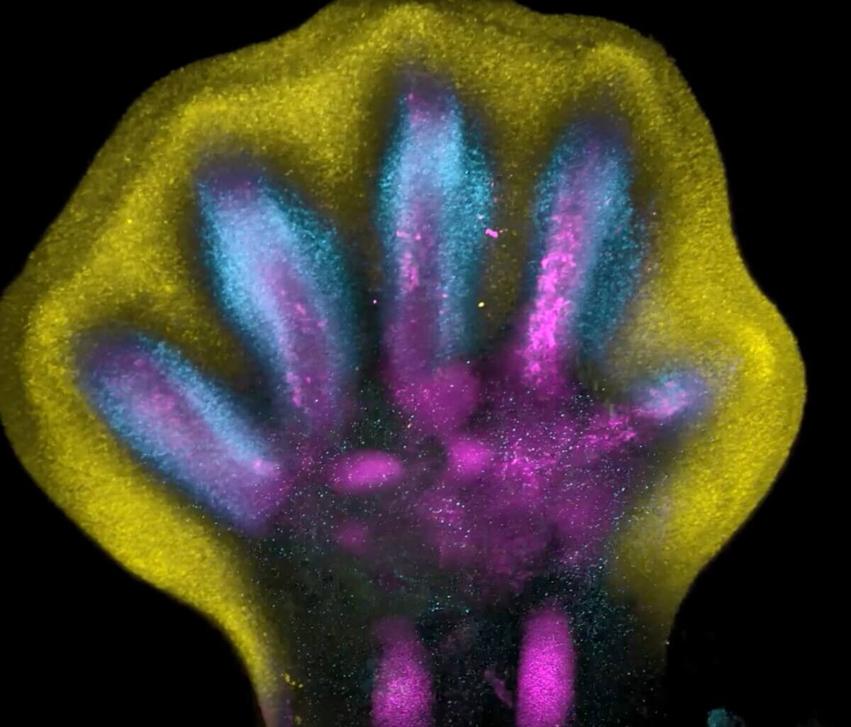
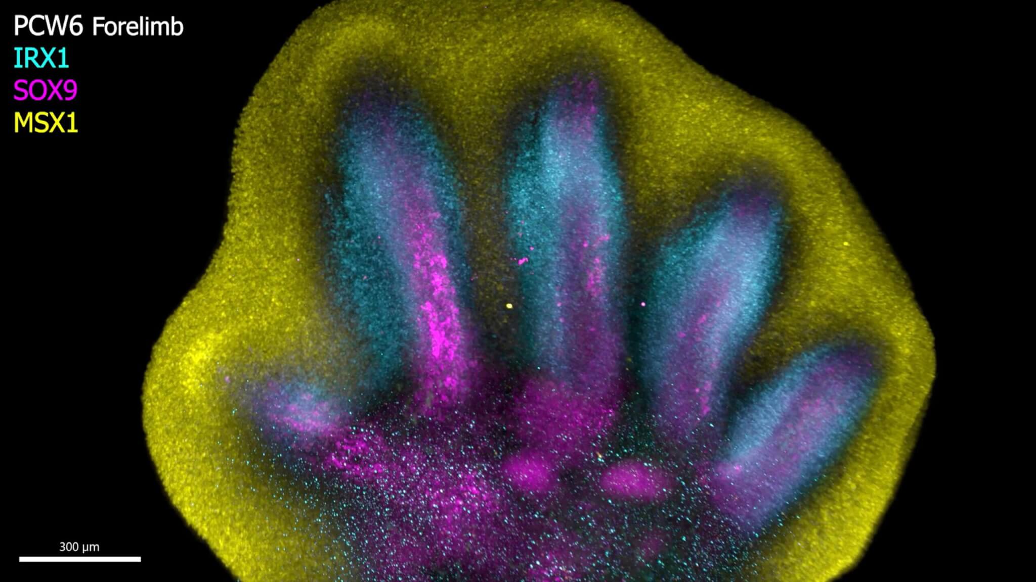
Still from video depicting how specific genes (IRX1, SOX9, and MSX1) work together to shape our fingers and toes during the development of limbs. IRX1 (blue) helps in forming our digits, SOX9 (purple) is important for building our bones, and MSX1 (yellow) is related to certain cells between the fingers and toes. Around the seventh week of development, some molecules cause specific cells in these spaces between fingers or toes to disappear. This process makes the distinct shapes of our fingers and toes visible. (Credit: SWNS)
HINXTON, United Kingdom — Scientists have unveiled groundbreaking insights into human limb development, providing new insights into the complex processes behind their formation. Unlike outward growth, human fingers and toes emerge from a larger foundational bud, with intervening cells receding to expose the digits. Approximately seven weeks into cell development, an orchestrated process of cell death reveals the distinct shapes of fingers and toes.
This discovery is part of a pioneering effort to create a spatial cell atlas of the entire developing human limb, capturing these processes for the first time. Using special tissue staining, researchers were able to observe how cell populations arrange themselves into the emerging patterns of digits.
The findings have significant implications for treating muscle-related disorders and injuries and may influence the diagnosis and treatment of congenital limb syndromes. Conducted as part of the Human Cell Atlas initiative, which aims to map every human cell type, the study involved researchers from the Wellcome Sanger Institute, Sun Yat-sen University, EMBL’s European Bioinformatics Institute, and other collaborators. They applied advanced single-cell and spatial technologies to chart the cellular landscape of the early human limb, pinpointing cell locations.
Published in the journal Nature, the atlas serves as an openly accessible resource, detailing the limbs’ rapid development during early formation. It also reveals new connections between developmental cells and some congenital limb syndromes, such as brachydactyly (short fingers) and polysyndactyly (extra digits).
Initially, limbs start as undefined cell pouches on the body’s sides, transforming into anatomically complex, recognizable limbs with fingers and toes after eight weeks. This rapid, precise cellular orchestration is susceptible to disturbances, leading to limb variations, some of the most common birth syndromes, affecting about one in 500 births worldwide.
While previous studies on limb development primarily used mouse and chick models, their relevance to human development was uncertain. However, technological advancements now enable the exploration of human limb formation’s early stages.
In this study, scientists analyzed tissues from five to nine weeks of development, tracing specific gene expression patterns that shape limbs. They identified certain genes, whose disruption is linked to specific limb syndromes, such as brachydactyly and polysyndactyly. Additionally, the research confirmed similarities in limb development between humans and mice.
Overall, these insights offer a comprehensive understanding of human limb development and hold the potential to impact congenital limb syndrome diagnosis and treatment.
“Decades of studying model organisms established the basis for our understanding of vertebrate limb development. However, characterizing this in humans has been elusive until now, and we couldn’t assume the relevance of mouse models for human development, says Professor Hongbo Zhang, the senior author of the study from Sun Yat-sen University, in a media release.
“What we reveal is a highly complex and precisely regulated process. It is like watching a sculptor at work, chiseling away at a block of marble to reveal a masterpiece. In this case, nature is the sculptor, and the result is the incredible complexity of our fingers and toes.”
“For the first time, we have been able to capture the remarkable process of limb development down to single-cell resolution in space and time, adds Dr. Sarah Teichmann, senior author of the study from the Wellcome Sanger Institute and co-founder of the Human Cell Atlas.
“Our work in the Human Cell Atlas is deepening our understanding of how anatomically complex structures form, helping us uncover the genetic and cellular processes behind healthy human development, with many implications for research and healthcare. For instance, we discovered novel roles of key genes MSC andPITX1 that may regulate muscle stem cells. This could offer potential for treating muscle-related disorders or injuries.”
South West News Service writer Dean Murray contributed to this report.


