
(Photo by Phonlamai Photo on Shutterstock)
OAK BROOK, Ill. — Artificial intelligence (AI) has the potential to significantly change the medical field, again. A new study shows that it can effectively use data from low-dose lung CT scans to improve predictions about a person’s risk for death from lung cancer, cardiovascular disease, and other conditions.
Currently, the U.S. Preventive Services Task Force recommends annual lung screenings with low-dose chest CT scans for people between the ages of 50 and 80 with a high risk of lung cancer, such as longtime smokers. In addition to images of the lungs, the scans can also provide information about other structures within the chest.
“When we’re looking at the CT images, the primary focus is on identifying nodules suspicious for lung cancer, but there is much more anatomical information coded in the space, including information on body composition,” says study lead author Kaiwen Xu, a Ph.D. candidate in the Department of Computer Science at Vanderbilt University, in a media release.
Previously, Xu and colleagues have developed, tested, and publicly released an AI algorithm that automatically derives body composition measurements from lung screening CT scans. These measures capture the percentage of fat, bone, and muscle in the body. Instances of abnormal body composition like obesity and loss of muscle mass are associated with chronic health conditions like heart disease. In fact, studies have shown that body composition is useful in identifying risk and prognosis for cardiovascular disease and chronic obstructive pulmonary disease (COPD). In lung cancer therapy, it has been shown to impact survival and overall quality of life.
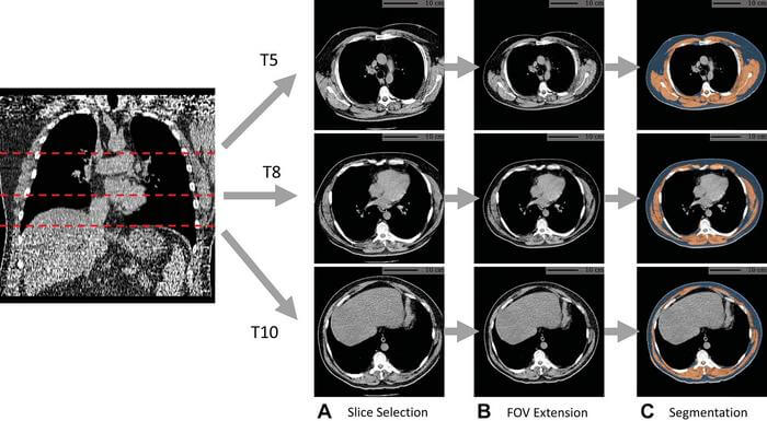
During this study, the team assessed the value added by AI body composition measurements by using CT scans of over 20,000 individuals from the National Lung Screening Trial. They found that by including these measurements, predicting lung cancer, heart disease, and all-cause mortality death risk was improved.
“Automatic AI body composition potentially extends the value of lung screening with low-dose CT beyond the early detection of lung cancer,” says Xu. “It can help us identify high-risk individuals for interventions like physical conditioning or lifestyle modifications, even at a very early stage before the onset of disease.”
Specifically, measurements linked to fat found in the muscle were especially strong mortality predictors. This particular finding is consistent with research already out there. Now, a condition called myosteatosis, which describes the infiltration of skeletal muscle with fat, is considered to be more predictive of health outcomes than a reduced muscle bulk.
These measurements go to show the imaging technique can be used for gathering information about other conditions, and not just give a snapshot of lung-specific ones. Therefore, integrating this more regularly within clinical care could be key.
“The images in a CT ordered for quite a different purpose—in our case, early detection of lung cancer—contain much more information,” Xu concludes. “In the space of the chest CT used for lung cancer screening, you can also check other information like body composition or coronary artery calcification that is directly associated with cardiovascular disease risk.”
The findings are published in the journal Radiology.


