(© adimas - stock.adobe.com)
ST. LOUIS, Mo. — Mindfulness is all about having peace of mind and body, even when it’s hard. Researchers from the Washington University School of Medicine in St. Louis now reveal just how intertwined the mind-body connection really is. They show that parts of the brain responsible for movement control are also closely linked to networks associated with thinking, planning, and even in blood pressure and heart control.
“People who meditate say that by calming your body with, say, breathing exercises, you also calm your mind,” says study first author Evan M. Gordon, PhD, an assistant professor of radiology at the School of Medicine’s Mallinckrodt Institute of Radiology.
“Those sorts of practices can be really helpful for people with anxiety, for example, but so far, there hasn’t been much scientific evidence for how it works. But now we’ve found a connection. We’ve found the place where the highly active, goal-oriented ‘go, go, go’ part of your mind connects to the parts of the brain that control breathing and heart rate. If you calm one down, it absolutely should have feedback effects on the other,” Gordon continues in a university release.
It all started in the 1930s, when neurosurgeon Wilder Penfield, MD, mapped motor areas of the brain using small jolts of electricity to the exposed part of the brain in people undergoing brain surgery to record their responses. He found that stimulating a narrow strip of tissue on each side of the brain causes specific body parts to twitch. He also found that control areas in the brain are arranged in the same order as the body parts they control — such as the toes being on one part of the strip while the face is on another. His map is now a neuroscience staple in several textbooks, referred to as the “little man” diagram.
Gordon and his team set out to replicate this work using functional magnetic resonance imaging (fMRI). They recruited seven healthy adults to undergo hours of fMRI brain scanning while resting or doing different tasks. They then built individualized brain maps for each participant, and validated their findings using the Human Connectome Project, the Adolescent Brain Cognitive Development Study, and the UK Biobank, which are publicly available datasets that have brain scans from around 50,000 people.
Researchers found errors in Penfield’s model they weren’t expecting
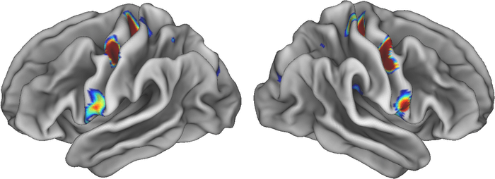
The control areas for feet, hands, and face were in the same spots, but there were three other areas that didn’t seem to be involved with movement at all, despite being in the motor region of the brain.
These non-movement areas looked different, too. They were thinner and strongly connected to each other as well as other parts of the brain involved in thinking, planning, mental arousal, pain, and control of functions like blood pressure. Although these areas weren’t activated by movement, they did become active when people thought about moving.
“All of these connections make sense if you think about what the brain is really for,” says senior author Nico Dosenbach, MD, PhD. “The brain is for successfully behaving in the environment so you can achieve your goals without hurting or killing yourself. You move your body for a reason. Of course, the motor areas must be connected to executive function and control of basic bodily processes, like blood pressure and pain. Pain is the most powerful feedback, right? You do something, and it hurts, and you think, ‘I’m not doing that again.’”
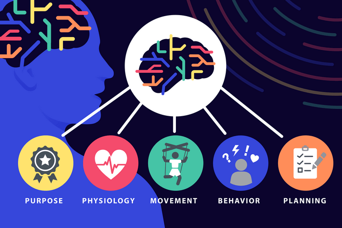
The team’s new network is called the Somato (body)-Cognitive (mind) Action Network, or SCAN, and they’ve so far analyzed the brains of a newborn, one-year-old, and nine-year-old. This work might help neuroscientists think about the mind-body connection in a whole new way.
“Penfield was brilliant, and his ideas have been dominant for 90 years, and it created a blind spot in the field,” explains Dosenbach. “Once we started looking for it, we found lots of published data that didn’t quite jibe with his ideas, and alternative interpretations that had been ignored. We pulled together a lot of different data in addition to our own observations, and zoomed out and synthesized it, and came up with a new way of thinking about how the body and the mind are tied together.”
The findings are published in the journal Nature.


