
Unconscious Young Man Lying On Floor In Living Room (© Andrey Popov - stock.adobe.com)
SAN DIEGO — What happens when people pass out? That question has stumped researchers for years, but a new study by a team at the University of California-San Diego has identified, for the very first time, the genetic pathway connecting the heart and brain in the context of fainting.
Fainting spells, also known as syncope, affect nearly 40 percent of people at least once in their lives. These episodes of momentary unconsciousness, triggered by various factors such as pain, fear, heat, or hyperventilation, contribute significantly to hospital emergency room visits. Prior to this latest research, the precise underlying mechanisms responsible for these “passing out” episodes have largely remained a mystery.
What sets this study apart is the novel perspective it brings to understanding this phenomenon. Instead of viewing the heart as a passive organ merely following the brain’s signals, the researchers explored the idea that the heart also sends signals back to the brain, influencing brain function.
“What we are finding is that the heart also sends signals back to the brain, which can change brain function,” says study senior author Vineet Augustine, assistant professor in the School of Biological Sciences at UCSD, in a university release.
This insight could have implications for understanding and treating various psychiatric and neurological disorders related to the connection between the brain and heart. The study, as stated in the paper, “is the first comprehensive demonstration of a genetically defined cardiac reflex, which faithfully recapitulates characteristics of human syncope at physiological, behavioral and neural network levels.”
The research delved into the neural mechanisms associated with the Bezold-Jarisch reflex (BJR), a cardiac reflex first described in 1867. For decades, researchers have speculated that the BJR, characterized by a decrease in heart rate, blood pressure, and breathing, might be linked to fainting. However, this hypothesis lacked substantial evidence due to limited knowledge of the neural pathways involved in the reflex.
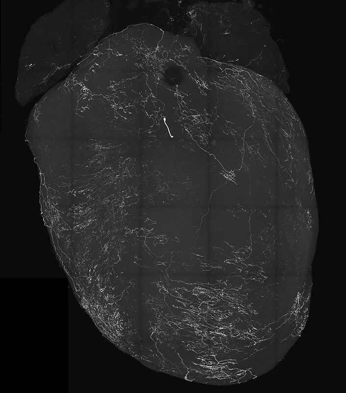
To uncover the genetics behind the sensory cluster known as the nodose ganglia, which are part of the vagus nerves responsible for transmitting signals between the brain and visceral organs, including the heart, the researchers focused on vagal sensory neurons (VSNs). They discovered that VSNs expressing the neuropeptide Y receptor Y2 (known as NPY2R) were tightly linked to the well-known BJR responses.
Using mice as their model, the researchers activated NPY2R VSNs using optogenetics, a method for stimulating and controlling neurons. The mice immediately fainted when these neurons were triggered. During these fainting episodes, researchers monitored thousands of neurons in the mice’s brains, heart activity, and changes in facial features, such as pupil dilation and whisker movement. They also employed machine learning to analyze the data, revealing rapid pupil dilation and the classic “eye-roll” observed during human fainting, along with suppressed heart rate, blood pressure, and breathing rate. The researchers also noted reduced blood flow to the brain.
“We were blown away when we saw how their eyes rolled back around the same time as brain activity rapidly dropped,” the researchers reported in a paper summary. “Then, after a few seconds, brain activity and movement returned. This was our eureka moment.”
Further experiments demonstrated that removing NPY2R VSNs from mice resulted in the disappearance of BJR and fainting conditions. The study not only confirmed that fainting is linked to reduced blood flow to the brain but also indicated that brain activity itself plays a crucial role. These findings implicate the activation of newly genetically identified VSNs and their neural pathways not only in BJR but also in overall animal physiology, specific brain networks, and behavior.
Such discoveries were challenging to uncover in the past because neuroscientists typically studied the brain, while cardiologists focused on the heart, often in isolation from each other.
“Neuroscientists traditionally think the body just follows the brain, but now it is becoming very clear that the body sends signals to the brain and then the brain changes function,” notes Augustine.
Based on their findings, researchers intend to further investigate the conditions that trigger vagal sensory neurons into action and study cerebral blood flow and neural pathways in the brain during syncope to gain a better understanding of this common yet enigmatic condition. Additionally, they hope to use their research as a model to develop targeted treatments for conditions associated with fainting.
The study is published in the journal Nature.
You might also be interested in:
- What happens when you die? Groundbreaking study first to record brain activity during death
- Theory of consciousness debunked: Key brain region actually acts like an internet router
- What happens to our consciousness when we fall asleep? Study may solve one of biggest scientific mysteries
Lea la versión en español en EstudioRevela.com: Misterio de los desmayos resuelto. El descubrimiento genético conecta el corazón y el cerebro cuando las personas se desmayan.


