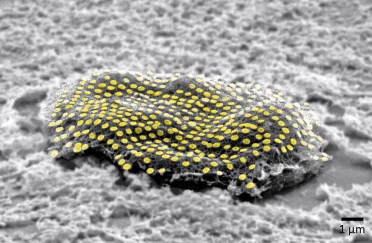
False-colored gold nanodot array on a fibroblast cell. (CREDIT: Kam Sang Kwok and Soo Jin Choi, Gracias Lab/Johns Hopkins University)
WASHINGTON — You won’t be able to go to a regular tattoo artist to get this type of ink. In a significant scientific advancement, Johns Hopkins University engineers have designed nanoscale tattoos that can adhere to living cells, bringing us one step closer to real-time monitoring of individual cell health.
This revolutionary technology introduces the ability to place optical components or electronics directly on live cells using “tattoo” arrangements. These arrangements seamlessly fit onto the cell’s wet and flexible exterior, much like a temporary tattoo on human skin.
“If you imagine where this is all going in the future, we would like to have sensors to remotely monitor and control the state of individual cells and the environment surrounding those cells in real time,” says David Gracias, a professor of chemical and biomolecular engineering at Johns Hopkins University who led the development of the technology, in a university release. “If we had technologies to track the health of isolated cells, we could maybe diagnose and treat diseases much earlier and not wait until the entire organ is damaged.”
Gracias likens these cellular tattoos to barcodes or QR codes, functioning as a link between living tissues and traditional sensors or electronic materials.
“We’re talking about putting something like an electronic tattoo on a living object tens of times smaller than the head of a pin,” notes Gracias. “It’s the first step towards attaching sensors and electronics on live cells.”
To ensure the tattoos stayed connected, they were designed using gold, a metal known for its conductive properties that prevent signal distortion. These were then attached to fibroblasts, cells responsible for tissue maintenance in humans. The bonding process involved molecular glues and an alginate hydrogel film, a gel-like substance that dissolves after ensuring the gold firmly sticks to the cell. The bond is further strengthened as the glue reacts with a film secreted by cells, known as the extracellular matrix.
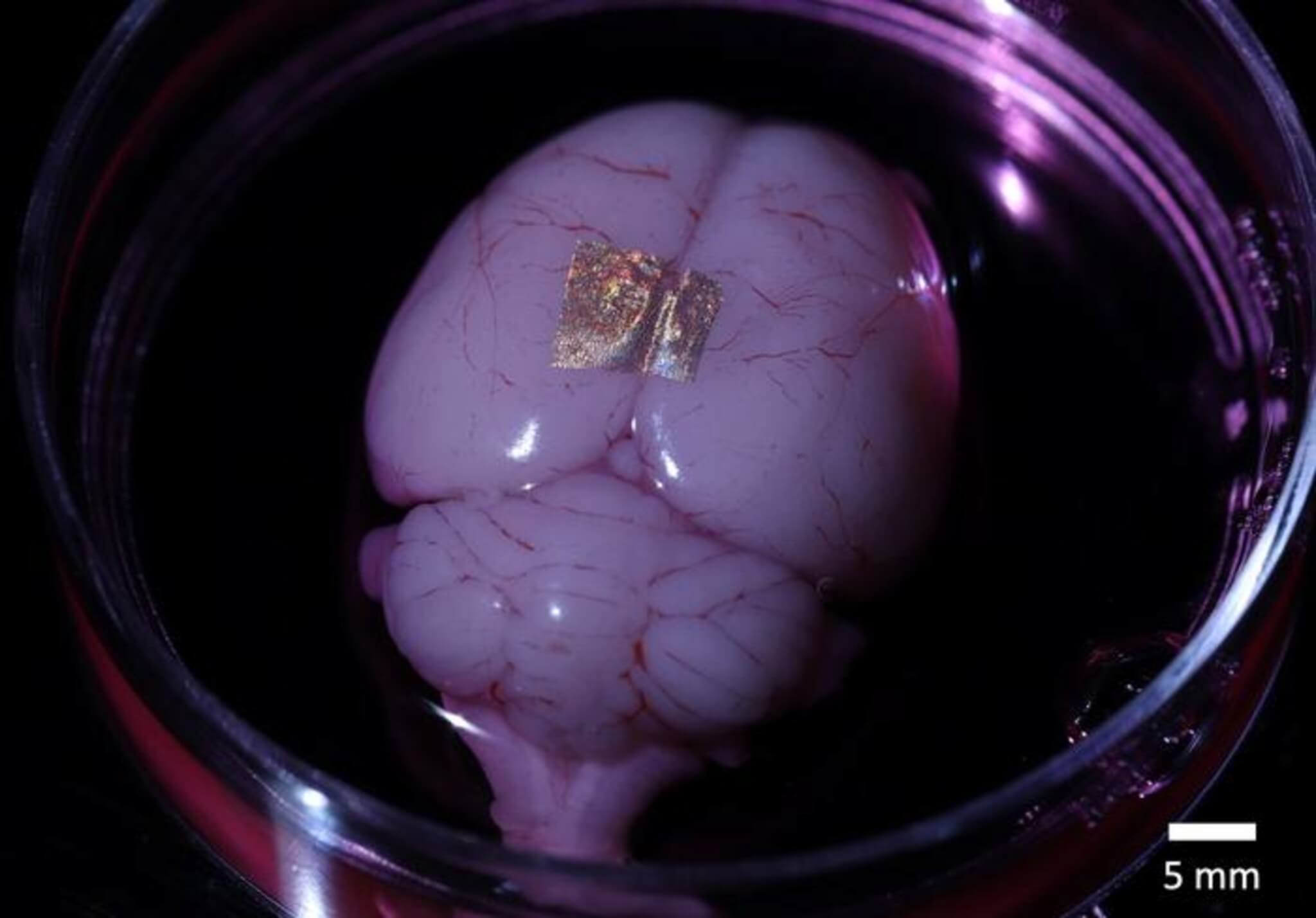
While past studies successfully applied hydrogels to stick nanotechnology onto human skin and animal organs, the team’s achievement lies in fixing nanowires and nanodots to individual cells. This addresses the historic difficulty of integrating optical sensors and electronics with biological matter on such a microscopic level.
“We’ve shown we can attach complex nanopatterns to living cells, while ensuring that the cell doesn’t die,” says Gracias. “It’s a very important result that the cells can live and move with the tattoos because there’s often a significant incompatibility between living cells and the methods engineers use to fabricate electronics.”
The strategic arrangement of these dots and wires is pivotal. For the technology to effectively track biological data, the sensors and wiring must be methodically placed, akin to electronic chip configurations.
“This is an array with specific spacing,” explains Gracias, “not a haphazard bunch of dots.”
The study is published in the journal Nano Letters.


Yeah, the study found what they were paid to find!