X-Ray illustration of brain stroke - 3d Illustration (Credit: Shutterstock)
OXFORD, United Kingdom — Scientists from the University of Oxford have developed an innovative 3D-printing technique that might one day revolutionize treatments for brain injuries. This technique involves creating neural cells that emulate the structure of the cerebral cortex, the brain’s outer layer.
Brain injuries, resulting from trauma, strokes, or surgeries for brain tumors, often lead to extensive damage to the cerebral cortex. This damage can hamper cognition, movement, and communication. A staggering 70 million people worldwide endure traumatic brain injuries annually, with five million of these instances being severe or even fatal. Presently, there is a dire absence of effective treatments for grave brain injuries, heavily compromising the quality of life for patients.
Potential solutions lie in tissue regenerative treatments, especially the kind where patients receive implants made from their own stem cells. The challenge has been ensuring these implanted stem cells accurately mirror the brain’s architecture.
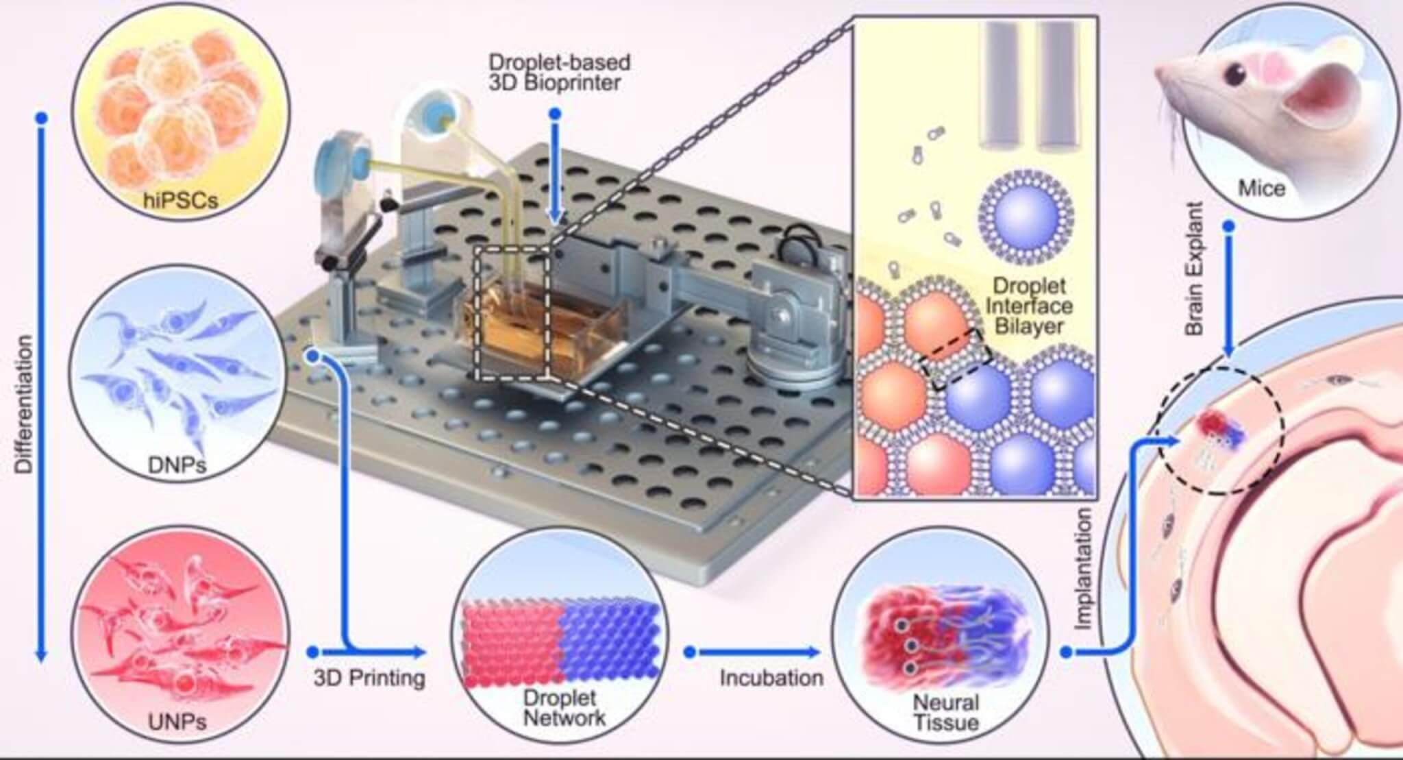
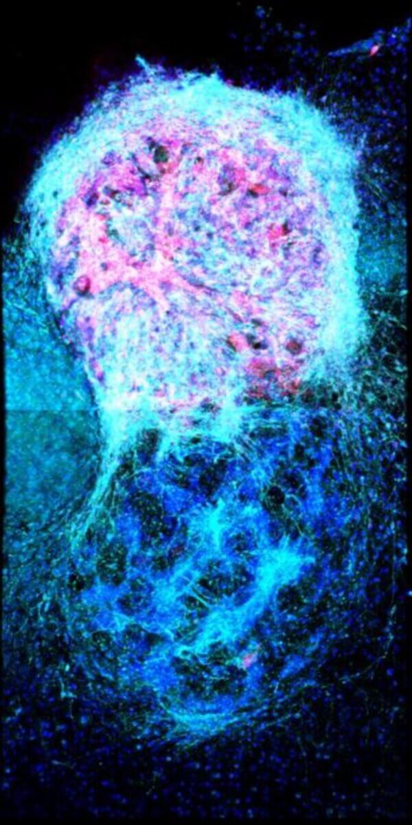
Diving deeper into this new approach, Oxford researchers constructed a two-layered brain tissue using 3D printing with human neural stem cells. These cells, when placed into mouse brain sections, merged seamlessly, both structurally and functionally, with the host tissue.
“This advance marks a significant step towards the fabrication of materials with the full structure and function of natural brain tissues,” says study lead author Dr. Yongcheng Jin, from Oxford’s Department of Chemistry, in a university release. “The work will provide a unique opportunity to explore the workings of the human cortex and, in the long term, it will offer hope to individuals who sustain brain injuries.”
These achievements were made possible using human induced pluripotent stem cells (hiPSCs). The cells can morph into most human tissue cell types. A vital benefit of hiPSCs for tissue repair is their origin: they can be derived from the patients themselves, ensuring no immune response when implanted.
By manipulating these hiPSCs with specific growth factors and chemicals, researchers created “bioinks” that were then 3D printed, maintaining their layered structure for weeks. When these printed tissues were introduced into mouse brains, they integrated deeply, proving both structural and functional coherence.
“Our droplet printing technique provides a means to engineer living 3D tissues with desired architectures, which brings us closer to the creation of personalized implantation treatments for brain injury,” notes study senior author Dr. Linna Zhou, from Oxford’s Department of Chemistry.
The research, which is a culmination of the team’s 10 years of experience in 3D printing technologies for synthetic tissues and cultured cells, showcases significant potential. Besides aiding brain injury repairs, this technology could also contribute to drug testing, studies on brain growth, and improving our grasp on cognition.
Researchers are now focusing on refining their technique, aspiring to recreate the intricate layers of the human brain’s architecture.
“Human brain development is a delicate and elaborate process with a complex choreography,” says study senior author Zoltán Molnár, a professor in the Department of Physiology, Anatomy and Genetics at Oxford. “It would be naïve to think that we can recreate the entire cellular progression in the laboratory. Nonetheless, our 3D printing project demonstrates substantial progress in controlling the fates and arrangements of human iPSCs to form the basic functional units of the cerebral cortex.”
The study is published in the journal Nature Communications.


