Non-cancerous breast conditions are far more common than breast cancer among women. Twenty percent of breast biopsies come back with a diagnosis of cancer. Look at it this way – there are hundreds of thousands of women with breast conditions that are not biopsied because they are not likely to be cancer. Add them to the equation and you get a tiny number of cases, far less than 20 percent, of breast changes that are malignant.
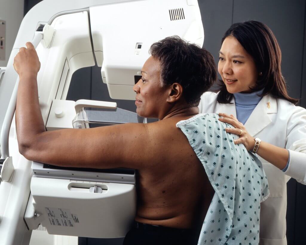
Women should not use that as an excuse to skip a mammogram. If you feel something in your breast that’s new, different, or painful, please see a doctor. It’s also a good idea to accept the testing or biopsy if your doctor recommends an in-depth examination. Sure, the chance that your breast findings are cancerous is smaller than small, but if breast cancer strikes, it’s important to catch it early.
However, let’s look at breast conditions that are not cancer, and whether they affect your risk for breast cancer as well.
Fibrocystic changes
Fibrosis, the root of the “fibro” in fibrocystic changes, refers to tough tissue similar to that of ligaments and scars. It can feel rubbery or firm. Cysts occur when fluid builds up in a breast gland. They can get as large as one to two centimeters. Types of cysts include the following:
- A simple cyst is filled with fluid. It does not increase your risk for cancer.
- A complicated cyst is like a simple cyst, but there is “debris” floating in the fluid. Complicated cysts are very unlikely to be cancer. In some cases, however, a doctor may advise additional examination. An ultrasound can distinguish between solid and fluid. A procedure to withdraw the fluid with a thin, hollow needle may be recommended to rule out cancer.
- A complex cystic and solid mass has thick outer walls or some solid tissue. These have a greater chance of being cancer than other types. A biopsy is usually necessary to determine if it is cancer (i.e., malignant) or non-cancerous (benign).
Fibrocystic changes are usually diagnosed when symptoms occur, such as lumps, swelling, tenderness, or pain. Symptoms tend to be worse just before a menstrual period.
Is there a treatment?
Fibrocystic changes in the breast are natural, so no treatment is necessary unless they are causing troublesome symptoms. Painful cysts can be drained using a needle and syringe. Drainage may reduce pressure and pain, but the fluid might come back later. Cysts may go away without treatment. Uncommonly, a surgeon removes recurring cysts.
For women with fibrocystic changes that don’t need treatment, a doctor might recommend watching the changes closely over time. Supportive bras, soothing heat, or over-the-counter (OTC) pain relievers, like acetaminophen (Tylenol) or ibuprofen (Motrin) relieve discomfort.
Some women say that their symptoms improve if they avoid caffeine and stimulant-containing products like coffee, tea, chocolate, and some soft drinks, although no scientific link between these stimulants and symptoms has been found. Many women feel that avoiding these foods and drinks is worth trying. Vitamins and herbal supplements are ineffective, and some have noxious, even dangerous, side-effects. Some doctors prescribe hormones for severe symptoms, but they can have serious side-effects as well.
Hyperplasia (ductal or lobular)
Hyperplasia is the overgrowth of the cells that line the milk-producing glands (lobules) or small tubes (ducts) within the breast. It is not cancer, but some types of hyperplasia increase the risk of developing breast cancer.
Hyperplasia can be described as either usual or atypical:
- Usual ductal hyperplasia causes an overgrowth of cells lining the ducts in the breast, but the cells look almost normal.
- Atypical hyperplasia produces abnormal cells. This can be either atypical ductal hyperplasia (ADH) or atypical lobular hyperplasia (ALH).
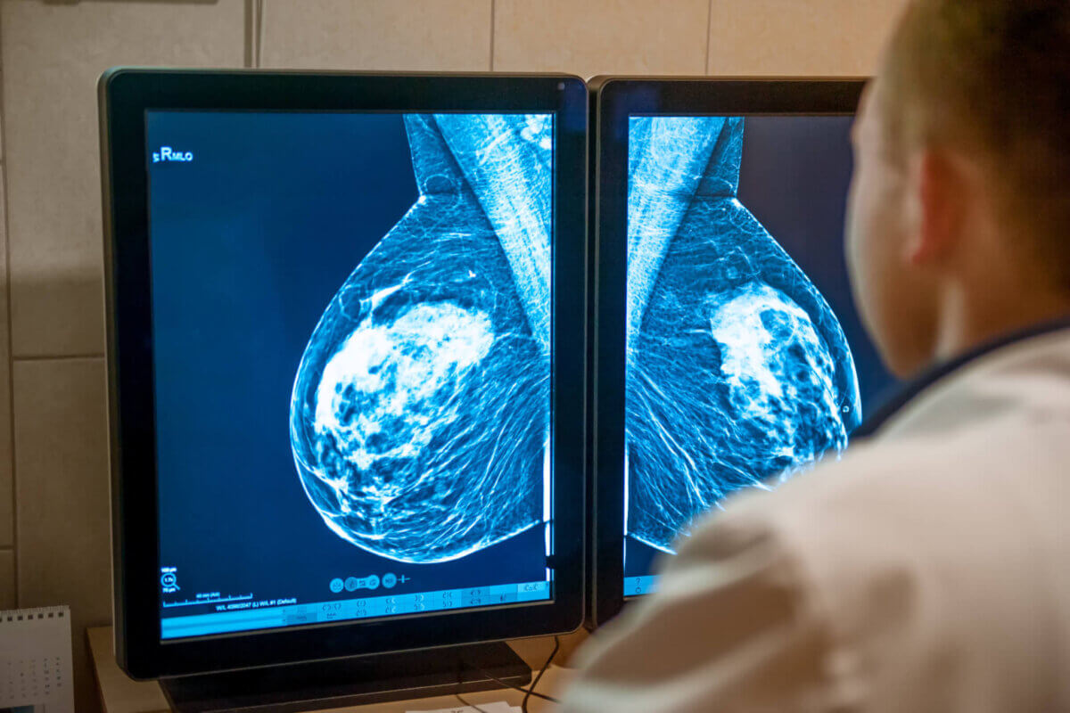
Hyperplasia does not usually cause a lump, but it may be seen on a mammogram. A biopsy is recommended, to remove some of the abnormal tissue for testing and exact diagnosis.
Hyperplasia can affect your risk for breast cancer – how much depends on what type of hyperplasia.
- Usual ductal hyperplasia increases the risk of breast cancer up to twice the risk of a woman with no breast abnormalities.
- Atypical hyperplasia (either ADH or ALH) increases the risk of breast cancer to about 4 to 5 times greater than a woman with no breast abnormalities.
Treatment of hyperplasia
Usual ductal hyperplasia does not need to be treated because it is a normal finding. If either ADH or ALH is found, surgery may be recommended to remove more breast tissue around the biopsy site. The tissue needs microscopic examination to be sure that there is no cancer nearby. If ADH or ALH without cancer is found after a surgical biopsy, typically no other treatment is needed.
Most women with ADH or ALH will not develop breast cancer. It’s still important, however, to talk with a healthcare provider about your risk and what you can do about it. Options may include:
- See a health care provider more often (such as every 6 to 12 months) for a breast exam along with a yearly mammogram. Breast magnetic resonance imaging (MRI) may be recommended, especially if you have other factors that raise your risk of breast cancer.
- Making lifestyle changes to lower breast cancer risk.
- Taking medicine to help lower breast cancer risk.
Adenosis
Adenosis is a benign condition in which some of the milk-producing glands of the breast (lobules) are enlarged. It’s often found along with fibrocystic changes. If many enlarged glands are found together, they may feel like a breast lump. Sometimes they contain calcifications that show up on mammograms. Similar calcifications may be seen with breast cancers, too. A breast biopsy is often necessary to distinguish breast cancers from adenosis.
Adenosis does not increase your risk for breast cancer and no treatment is needed.
Fibroadenomas
Fibroadenomas are common, benign breast tumors made up of both glandular and connective tissues. They are most common in women in their 20s and 30s but can be found in women of any age. They tend to shrink after menopause. Fibroadenomas are often round or oval and may feel like a marble within the breast tissue. They move under the skin and are not tender. Small fibroadenomas may be found on ultrasound or mammograms. They can become so large that they deform the shape of the breast.
Fibroadenomas do not increase your risk for breast cancer. No treatment is needed. If they are not growing, they can be left in place. At times, surgical removal would have to include significant amounts of normal breast tissue, deforming the breast.
Lobular carcinoma in situ (LCIS)
The name lobular carcinoma in situ (LCIS) sounds frightening but the condition is not cancerous. It usually doesn’t spread beyond the lobules. It is an incidental finding on microscopic examination of tissue from a breast biopsy done for some other problem nearby in the breast. Cells that look like cancer cells are growing in the lining of the milk-producing glands. The different types of LCIS are:
- Classic LCIS shows cells lining the lobules of the breast which are smaller than normal cells.
- Pleomorphic LCIS shows cells lining the lobules of the breast are larger than normal and look abnormal.
- Florid LCIS consists of the cells lining the lobules growing into a large enough group that they have formed a mass, usually with some dead cells in the middle of the mass (called central necrosis).
Women with LCIS have a higher risk of developing invasive cancer in either breast – seven to 12 times the risk of a woman with normal breast tissue. Doctors usually recommend that women with LCIS have regular breast cancer screening tests and visits with a healthcare provider every six to 12 months for the rest of their lives.
What’s the treatment for LCIS?
Although LCIS increases your risk of developing invasive breast cancer, it is not cancer or pre-cancer, so treatment is often not necessary after the biopsy.
Sometimes if LCIS is found using a needle biopsy, the doctor might recommend that it be removed completely (with an excisional biopsy or some other type of breast-conserving surgery) to help make sure that LCIS is the only abnormality. This is especially true if the LCIS is described as pleomorphic or florid, in which case it might be more likely to grow quickly. Even after an excisional biopsy, if pleomorphic or florid LCIS is found, some doctors might recommend another, more extensive surgery to make sure it has all been removed.
Close follow-up is important because women with LCIS have the same increased risk of developing cancer in both breasts. Women should also talk to a healthcare provider about what they can do to help reduce their breast cancer risk. Options for women at high risk of breast cancer because of LCIS may include:
- See a healthcare provider more often (such as every 6 to 12 months) for a breast exam along with a yearly mammogram. Additional imaging with breast MRI may also be recommended, especially if a woman has other factors that raise her risk of breast cancer.
- Making lifestyle changes to lower breast cancer risk.
- Taking medicine to help lower the risk of breast cancer.
- Surgery, called bilateral prophylactic mastectomy (removal of both breasts), to reduce risk. This is more likely to be a reasonable option in women who also have other risk factors for breast cancer.
If something about your health triggers the thought, “I wonder if I should see my healthcare provider?” the answer is yes.
Sources:
- The American Cancer Society
- The Mayo Clinic
- The Cleveland Clinic
- The Centers for Disease Control and Prevention