In hopes of improving the survival rate for breast cancer patients, MIT researchers designed a wearable ultrasound device that could allow women to detect tumors when they are still in early stages. (credit: Canan Dagdeviren)
CAMBRIDGE, Mass. — A new wearable ultrasound patch designed to fit inside a bra could expedite the detection of breast cancer. This device is engineered to monitor breast tissue and identify tumors in their early stages, significantly boosting patients’ survival rates.
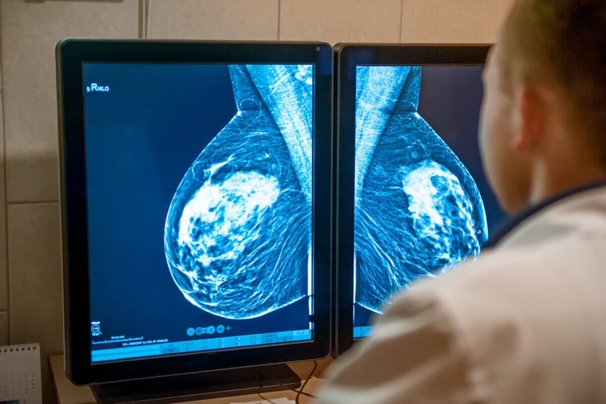
The invention comes from American researchers aiming to combat “interval cancers,” which emerge between regular scans and constitute almost 30 percent of all breast cancer cases. The survival rate for breast cancer – the most common cancer in women – is nearly 100 percent when diagnosed early. However, that figure plummets to just 25 percent for tumors detected in later stages.
The research team crafted this wearable device as a home monitoring method for those at high risk of breast cancer. The study’s lead author, Turkish scientist Dr. Canan Dağdeviren, an Associate Professor at the Massachusetts Institute of Technology’s Media Lab, is personally affected by the tragic loss of her aunt to late-stage breast cancer, was inspired to create a wearable diagnostic device that could be incorporated into a bra.
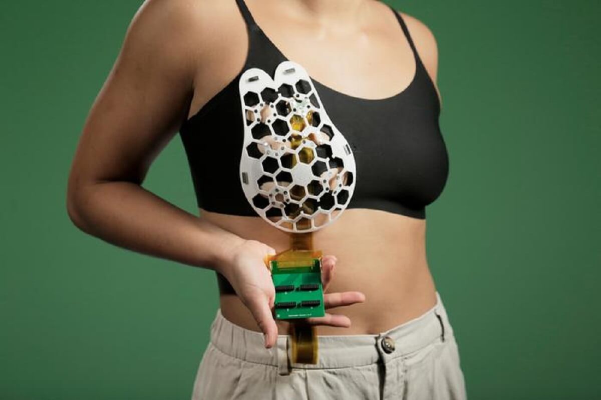
The resulting ultrasound device aims to facilitate earlier breast cancer diagnoses in women. The device was proven to capture images as accurately as those taken at medical imaging centers. The team designed it as a flexible patch that attaches to a bra, allowing the wearer to move freely while an ultrasound tracker captures images of breast tissue from different angles.
“We changed the form factor of the ultrasound technology so that it can be used in your home. It’s portable and easy to use, and provides real-time, user-friendly monitoring of breast tissue,” says Dr. Dağdeviren in a media release. “My goal is to target the people who are most likely to develop interval cancer. With more frequent screening, our goal is to increase the survival rate to up to 98 percent.”
Interval cancers, or tumors that develop between scheduled mammogram breast screenings, account for 20 to 30 percent of all breast cancer cases and tend to be more aggressive than those detected during routine scans.
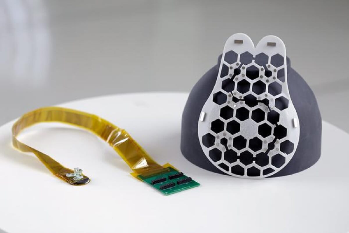
To make the device wearable, the team devised a flexible, 3D-printed patch with honeycomb-like openings. Using magnets, it attaches to a bra, allowing the scanner to contact the wearer’s skin. The ultrasound scanner is housed inside a small tracker, which can move to six different positions, allowing for complete breast imaging. It can also rotate to capture images from different angles and doesn’t require specialist expertise to operate.
The device was tested on a 71-year-old woman with a history of breast cysts and could detect cysts as small as 0.3 centimeters in diameter, similar in size to early-stage tumors.
“Access to quality and affordable health care is essential for early detection and diagnosis,” says Catherine Ricciardi, nurse director at MIT’s Center for Clinical and Translational Research. “This technology holds the promise of breaking down the many barriers for early breast cancer detection by providing a more reliable, comfortable, and less intimidating diagnostic.”
Though wearers currently have to connect their scanners to a traditional ultrasound machine, the research team is working on a miniaturized imaging system about the size of a smartphone. The ultrasound patch can be reused, and the researchers envision it being used at home by those at high risk of breast cancer and those without access to regular screening.
The team also plans to incorporate advances in artificial intelligence to track how images change over time, offering more precise diagnostics. They also aim to adapt the technology to scan other body parts for early diagnoses of other cancers.
The study is published in the journal Science Advances.
South West News Service writer James Gamble contributed to this report.


Spectacular.
Science is now rapidly gaining over all forms of disease.
Religion however, has done nothing over the last 4,000 yearss.
Check you bias. Do you hate religion so much that you deny it was an unavoidable requirement of our evolution towards science and critical thought? People had to develop language, writing, and societal structure before we could have ever reached the science of today. Religion is part of our history, it was the human mind imagining how things worked before it had the ability to understand or measure reality. Religion will be left behind as science advances, but to make a statement saying it did nothing to benefit us 4000 years ago is truly ignorant to the struggle humanity had to push toward to get to today’s understanding.
Careful folks? Sounds like a godsend, but we’ve seen this movie before. Haven’t we? First, you’re told, it helps with early detection. Years from now, we may be told, … Oops!! It’s become a contributing factor. Same old song.
How about skipping the bra instead, which is the best way to prevent breast cancer. I am a breast cancer researcher and co-author of Dressed to Kill: The Link Between Breast Cancer and Bras. Studies from around the world now confirm the bra-cancer link. Bra-free women have about the same risk of breast cancer as men, while the longer and tighter the bra is worn the higher the risk rises, to over 100 times higher for a 24/7 bra wearer compared to a bra-free woman. If a bra leaves marks in the skin, it is too tight and is harming the breasts.