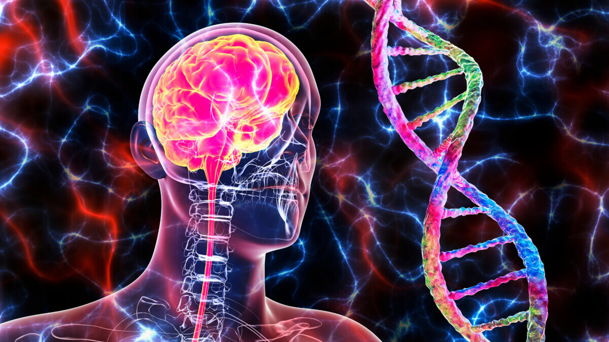
Genetic brain disorders (© Dr_Microbe - stock.adobe.com)
NEW YORK — Have you ever wondered what happens to your brain after you die? Scientists at the Icahn School of Medicine at Mount Sinai have just uncovered some mind-blowing differences between living and dead brain tissue, and it’s shaking up everything we thought we knew about the human brain!
Discovering the Brain’s Secret Language
In a remarkable leap forward for neuroscience, researchers discovered stark differences in RNA editing between living and postmortem brains. Specifically, this pioneering research, published in Nature Communications, highlights the nuances of adenosine-to-inosine (A-to-I) RNA editing in the brain’s dorsolateral prefrontal cortex (DLPFC). So, what exactly does that mean?
First, let’s talk about “RNA editing.” Don’t worry if it sounds like science jargon – it’s actually quite interesting! Imagine your genes as a blueprint for your body. RNA editing is like a master editor that can tweak that blueprint on the fly, allowing your brain to adapt and function smoothly.
Scientists have been studying this process for years, but with a catch: they’ve mostly been looking at brain tissue from people who have passed away. It turns out that might not tell the whole story.
“Until now, the investigation of A-to-I editing and its biological significance in the mammalian brain has been restricted to the analysis of postmortem tissues. By using fresh samples from living individuals, we were able to uncover significant differences in RNA editing activity that previous studies, relying only on postmortem samples, may have overlooked,” explains Dr. Michael Breen, the co-senior author of the study in a media release.
“We were particularly surprised to find that RNA editing levels were significantly higher in postmortem brain tissue compared to living tissue, which is likely due to postmortem changes such as inflammation and hypoxia that do not occur in living brains. Additionally, we discovered that RNA editing in living tissue tends to involve evolutionarily conserved and functionally important sites that are also dysregulated in human disease, emphasizing the need to study both living and postmortem samples for a comprehensive understanding of brain biology.”

Methodology: Examining the Living and Dead
In the study, researchers compared brain samples from living people who volunteered to be examined during brain surgery with samples from those who had died. These samples were meticulously preserved and analyzed using advanced genomic techniques, including whole-genome sequencing (WGS), bulk-tissue RNA sequencing (RNA-seq), and single nuclei RNA sequencing (snRNA-seq).
Key Results: A Tale of Two Brains
The dead brain tissue showed much more RNA editing than the living tissue. It’s like the brain goes into overdrive after death, making changes left and right. But why does this matter?
For years, scientists have been using postmortem brain tissue to study everything from Alzheimer’s to depression. However, if the brain changes so much after death, it means we might have been getting an incomplete picture all along. Think of it like studying a city after a major earthquake. Sure, you can learn some things, but you’re not seeing how the city normally functions. That’s the challenge scientists are now facing with brain research.
“Understanding these differences helps improve our knowledge of brain function and disease through the lens of RNA editing modifications, which can potentially lead to better diagnostic and therapeutic approaches,” notes Alexander W. Charney, MD, PhD, from the Living Brain Project.
Takeaways: Changing the Future of Brain Science
This discovery is opening up a whole new world of possibilities. Researchers are now looking for ways to get more samples from living brains safely and ethically. They’re also working on developing new techniques to preserve brain tissue better after death, as well as combining data from both living and deceased brains for a fuller picture.
While it might sound like a far-off scientific debate, this research could have a huge impact on how we understand and treat brain diseases. By getting a clearer picture of how healthy brains work, especially before death, scientists might find new ways to tackle conditions like Parkinson’s, Alzheimer’s, and depression.
“By harnessing the unique, transdisciplinary nature of the Living Brain Project, we can turn a cutting edge clinical care modality like deep brain stimulation into a platform for unprecedented insight into human brain biology that will give rise to new therapeutic opportunities,” concludes Brian Kopell, MD, co-first author of the study.


