(© ra2 studio - stock.adobe.com)
LONDON — Going to the eye doctor could help detect Parkinson’s disease up to seven years before experts make an official diagnosis. Researchers at University College Hospital and the Moorfields Eye Hospital say this eye test represents the first instance where such early detection has been possible. The study is the most extensive examination of retinal imaging in relation to Parkinson’s disease, a debilitating neurodegenerative condition impacting approximately one million individuals in the United States alone.
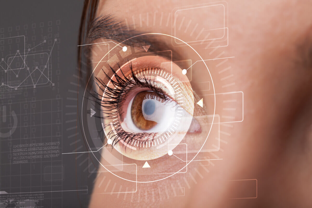
To find markers of Parkinson’s in eye scans, the research team turned to artificial intelligence (AI), utilizing the AlzEye dataset. They validated their findings by cross-referencing with the extensive UK Biobank database of healthy patients. By leveraging these robust datasets, the scientists could pinpoint nuanced markers, even though Parkinson’s disease only affects a minute percentage of the population.
The research underscores the growing role of eye scans, or “oculomics,” in disease detection. Over recent years, this method has proven effective in identifying other neurodegenerative conditions such as Alzheimer’s, multiple sclerosis, and even schizophrenia. Eye scans have also been instrumental in highlighting risks for high blood pressure, and cardiovascular diseases including strokes and diabetes.
Parkinson’s disease is typified by diminished dopamine levels. Autopsies of Parkinson’s patients have unveiled discrepancies in the retina’s inner nuclear layer (INL). A noticeable thinning of this layer has been correlated with a heightened probability of the disease’s onset.
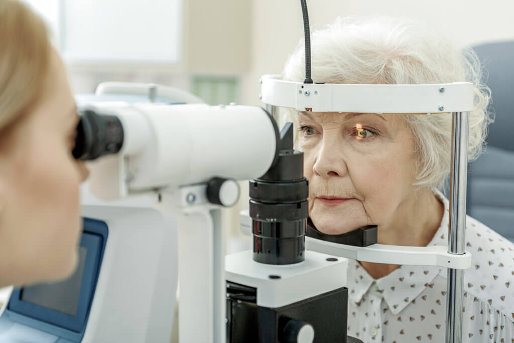
Today, high-resolution retinal imaging, especially through “optical coherence tomography” (OCT), has become a standard in eye care. An OCT scan offers a detailed cross-section of the retina, producing images with precision up to a thousandth of a millimeter. Leveraging advanced computational power, scientists can swiftly analyze vast quantities of OCTs, with machine learning revealing comprehensive insights about the human body from these scans.
“I continue to be amazed by what we can discover through eye scans,” says lead author Dr. Siegfried Wagner of the UCL Institute of Ophthalmology and Moorfields Eye Hospital, in a media release. “While we are not yet ready to predict whether an individual will develop Parkinson’s, we hope that this method could soon become a pre-screening tool for people at risk of disease. Finding signs of a number of diseases before symptoms emerge means that, in the future, people could have the time to make lifestyle changes to prevent some conditions arising, and clinicians could delay the onset and impact of lifechanging neurodegenerative disorders ”
This research reaffirms previous findings regarding the thinning of the ganglion cell–inner plexiform layer (GCIPL) while, for the first time, observing a reduced INL. Future studies will aim to discern if GCIPL atrophy progression is tied to brain alterations in Parkinson’s or if INL thinning is a precursor.
Understanding this dynamic could provide a deeper comprehension of the disease’s triggers, and determine if retinal imaging can bolster the diagnosis, prognosis, and comprehensive patient management in Parkinson’s cases.
“Broadening the scope of imaging across a larger segment of the population could profoundly benefit public health,” emphasizes Moorfields’ medical director, Louisa Wickham. “Increasing imaging across a wider population will have a huge impact on public health in the future, and will eventually lead to predictive analysis. OCT scans are more scalable, non-invasive, lower cost and quicker than brain scans for this purpose.”
The study is published in the journal Neurology.
You might also be interested in:
- AI identifies 4 types of Parkinson’s disease, paving way for personalized treatments
- Could a common eye test detect Alzheimer’s disease decades earlier?
- Potential breakthrough treatment created for Alzheimer’s, Parkinson’s disease
South West News Service writer Jim Leffman contributed to this report.


