
Photo by KeithJJ from Pixabay
CHICAGO — Much has been made in recent years regarding the risk of brain injuries tied to American football and other contact sports. Now, noteworthy new findings suggest soccer, or European football, may also expose athletes to a serious risk of head injuries. Specifically, soccer players may endanger their brain function by heading the ball often.
Findings reveal a connection between soccer heading (when a player hits the ball with their head) and a measurable decline in the microstructure and function of the mind over a two-year period.
“There is enormous worldwide concern for brain injury in general and in the potential for soccer heading to cause long-term adverse brain effects in particular,” says senior author Michael L. Lipton, M.D., Ph.D., professor of radiology at Columbia University’s Vagelos College of Physicians and Surgeons and affiliate professor of biomedical engineering at Columbia University, in a media release. “A large part of this concern relates to the potential for changes in young adulthood to confer risk for neurodegeneration and dementia later in life.”
Prior studies have analyzed the adverse effects on the brain related to soccer heading at singular points in time, but this latest project opted to focus on brain changes over two years. In all, the study included 148 young adult amateur soccer players with an average age of 27 years old and 26 percent being women. Researchers put together a specialized questionnaire for players aimed at determining how often they hit the soccer ball with their heads.
“When we first started, there was no method for assessing the number of head impacts a player experienced,” Dr. Lipton comments. “So, we developed a structured, epidemiological questionnaire that has been validated in multiple studies.”
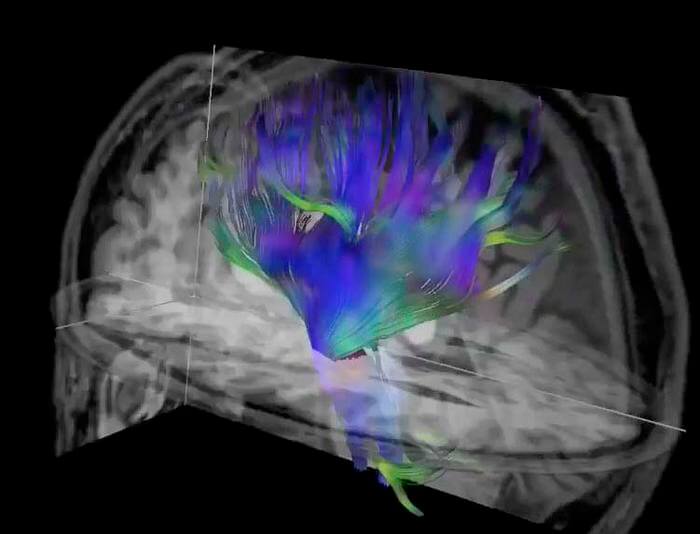
The survey featured a series of questions about how often the individual played, practiced, headed the ball, and in what type of situations. Two-year heading exposure was categorized as low, moderate, or high. Players were also analyzed for verbal learning and memory and then underwent diffusion tensor imaging (DTI), an MRI technique, at the time of enrollment and then once again two years later. DTI can characterize the microstructure of the brain through the tracking of microscopic movements of water molecules through the tissue.
In comparison to the baseline test results, the high-heading group (over 1,500 headers in two years) showed a clear increase of diffusivity in frontal white matter regions and a notable decrease in orientation dispersion index (a measure of brain organization) across certain brain regions after two years of heading exposure. This held true even after researchers accounted for numerous variables such as age, sex, education, and concussion history.
“Our analysis found that high levels of heading over the two-year period were associated with changes in brain microstructure similar to findings seen in mild traumatic brain injuries,” Dr. Lipton adds. “High levels of heading were also associated with a decline in verbal learning performance. This is the first study to show a change of brain structure over the long term related to sub-concussive head impacts in soccer.”
Dr. Lipton and his colleagues also conducted a second study in which they used DTI to investigate the association between repetitive head impacts from soccer heading and verbal learning performance. During this project, the researchers analyzed heading for 12 months prior to DTI and verbal learning performance testing in 353 amateur soccer players between the ages of 18 and 53. However, unlike earlier studies that had focused on deep white matter regions, this study employed a new technique. The team utilized DTI parameters to assess the integrity of the interface between the brain’s gray and white matter very close to the skull.
“Importantly, our new approach addresses a brain region that is susceptible to injury but has been neglected due to limitations of existing methods,” Dr. Lipton says. “Application of this technique has potential to disclose the extent of injury from repetitive heading, but also from concussion and traumatic brain injury to an extent not previously possible.”

Ultimately, researchers found that the normally sharp gray matter-white matter interface became blunted in proportion to high repetitive head impact exposure.
“We used DTI to assess the sharpness of the transition from gray matter to white matter,” Dr. Lipton notes. “In various brain disorders, what is typically a sharp distinction between these two brain tissues becomes a more gradual, or fuzzier transition.”
He adds that gray matter-white matter interface integrity could play a causal role in the adverse association connecting repetitive head impacts and cognitive performance.
“These findings add to the ongoing conversation and contentious debate as to whether soccer heading is benign or confers significant risk,” the researcher concludes.
This research was presented at the 109th Scientific Assembly and Annual Meeting of the Radiological Society of North America.


