NASHVILLE, Tenn. — Vanderbilt University researchers have made a startling discovery in the human brain — recording a forceful and unexplained signal in the brain’s white matter.
The human brain is composed of two distinct types of matter: gray matter and white matter. Gray matter, home to nerve cell bodies, is responsible for processing sensation, controlling voluntary movement, and enabling speech, learning, and cognition. On the other hand, white matter serves as a vast network of connections, linking cells to each other and projecting signals throughout the body.
Scientists have predominantly focused their attention on the gray matter of the brain, considering it the hub of activity while overlooking the significance of white matter, despite it constituting half of the brain’s composition. Researchers at Vanderbilt University are determined to change this narrative.
For several years, a team led by Dr. John Gore, director of the Vanderbilt University Institute of Imaging Science, has been employing functional magnetic resonance imaging (fMRI) to detect blood oxygenation-level dependent (BOLD) signals, a critical indicator of brain activity, within the brain’s white matter.
In their latest study, researchers have revealed a remarkable finding: when individuals undergoing brain scans using fMRI perform tasks such as finger wiggling, BOLD signals in white matter across the entire brain increase.
“We don’t know what this means,” says study first author Dr. Kurt Schilling, research assistant professor of Radiology and Radiological Sciences at VUMC, in a university release. “We just know that something is happening. There truly is a powerful signal in the white matter.”

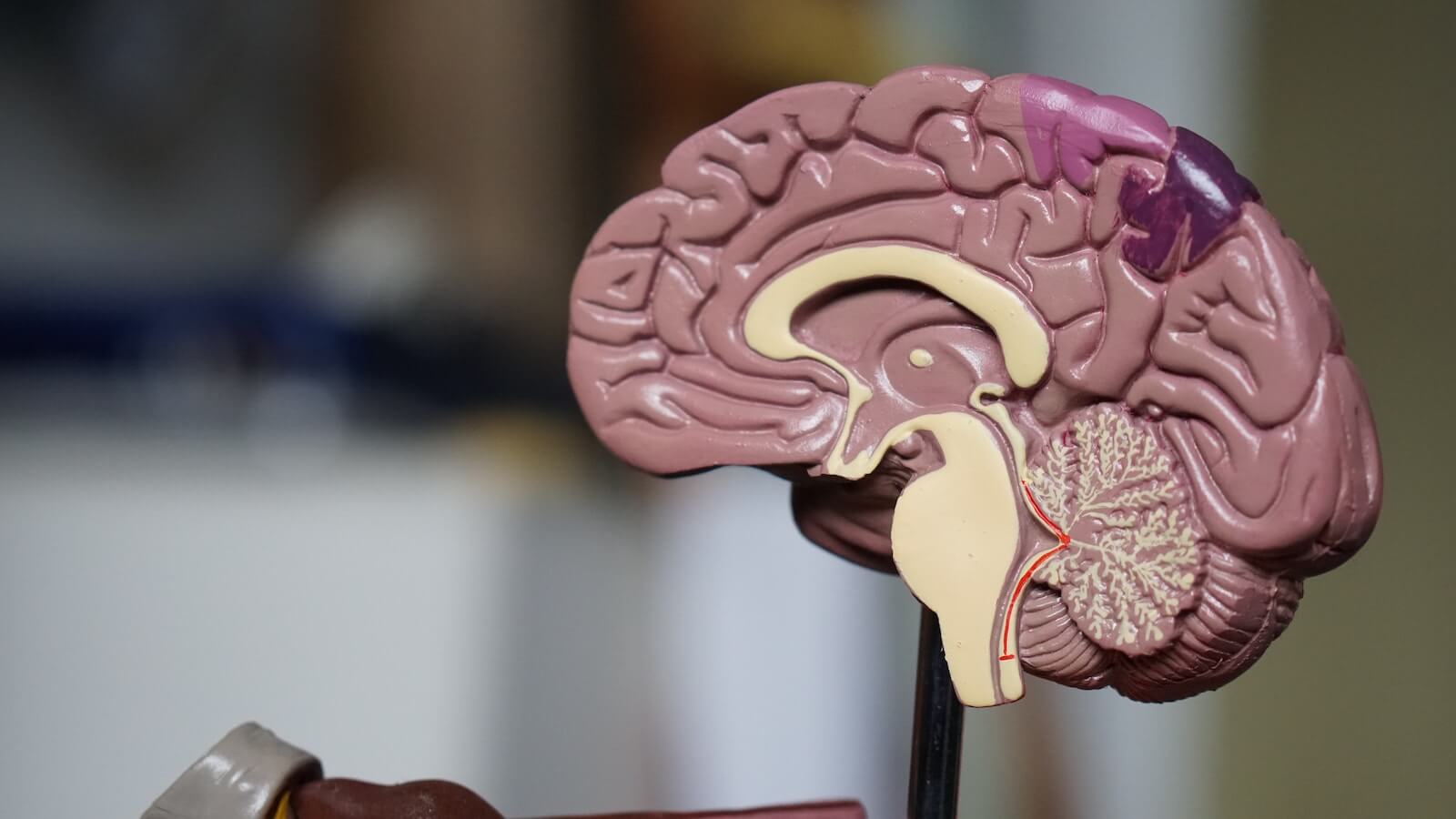
(credit: Photo by Robina Weermeijer on Unsplash)
This discovery is significant because various disorders, including epilepsy and multiple sclerosis, disrupt the brain’s “connectivity.” This suggests that white matter plays a crucial role in these conditions, prompting the need for further investigation.
To unravel the mystery, researchers plan to continue examining changes in white matter signals previously observed in conditions like schizophrenia and Alzheimer’s disease. Through animal studies and tissue analysis, they aim to uncover the biological underpinnings of these changes.
In gray matter, BOLD signals signify an increase in blood flow and oxygen consumption in response to heightened nerve cell activity. It remains to be seen whether axons (long projections of nerve cells) or the glial cells responsible for maintaining the protective myelin sheath around them also consume more oxygen during brain activity. Alternatively, these signals may be linked to what’s transpiring in the gray matter.
Even if there’s no biological activity occurring in the white matter, “there’s still something happening here,” notes Schilling. “The signal is changing. It’s changing differently in different white matter pathways and it’s in all white matter pathways, which is a unique finding.”
One of the reasons white matter signals have received less attention is their lower energy compared to gray matter signals, making them harder to distinguish from the background “noise” in brain scans. Vanderbilt scientists overcame this challenge by having individuals undergoing brain scans repeat visual, verbal, or motor tasks multiple times to establish patterns and by averaging the signals across various white matter pathways.
“For 25 or 30 years, we’ve neglected the other half of the brain,” says Schilling.
In some cases, researchers not only overlooked white matter signals but also omitted them from their reports on brain function.
The Vanderbilt study implies that numerous fMRI studies may not only underestimate the full extent of brain activation but also potentially miss vital information conveyed by MRI signals, ultimately opening new avenues of exploration in neuroscience.
The study is published in the journal Proceedings of the National Academy of Sciences.
You might also be interested in:
- What happens after death? Scientists discover ‘surge of activity’ in the dying brain
- Brain activity much lower using Zoom than during in-person conversations, study reveals
- Mystery: Brain naturally produces powerful psychedelic compound DMT, study finds


Ever feel someone’s energy is off? This will be proven to be how we communicate noverbally through energy. Just you watch!
I was told 30 years ago when I had a MRI that my white matter had ‘hyperactivity’. Still have no idea what it means. Now, at 61, other than typical men issues, I feel fine! Retired 5 years now, I go when and where I can. Life is to short. I’m anxious to see where this and future white matter activity reports go.