CAMBRIDGE, Mass. — A cutting-edge ultrasound patch capable of determining bladder fullness has potential future applications in early cancer detection, scientists say.
This wearable monitor, designed for individuals with bladder and kidney conditions, allows for internal organ imaging without the need for a gel application or an ultrasound technician. Researchers at the Massachusetts Institute of Technology (MIT) are optimistic that the device can be modified to monitor other organs, aiding in the early identification of various cancers.
The device was conceived by Dr. Canan Dağdeviren, an associate professor at MIT’s Media Lab and the study’s senior author, who was motivated by her brother’s struggle with kidney cancer. Following the removal of one kidney, he had difficulties with bladder emptiness, which prompted Dr. Dağdeviren to focus the patch’s initial applications on bladder monitoring.
The lab, which has a history of developing flexible, wearable electronics, recently invented an ultrasound monitor for potential integration into a bra for breast cancer screening.
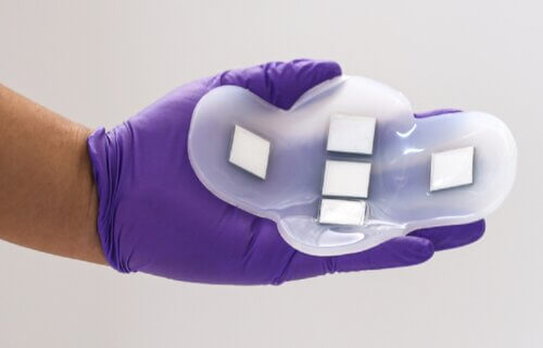
For the latest study, the team created a wearable device using a new “piezoelectric” material in a flexible silicone rubber patch with five ultrasound arrays to take pictures of the bladder and other internal organs, assisting individuals like her brother in monitoring their bladder fullness without visiting a medical facility.
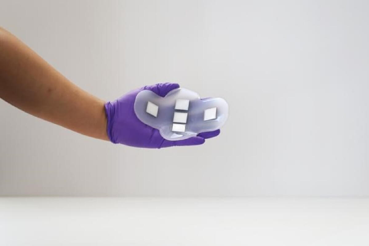
(Credit: MIT)
Traditional bladder volume measurement necessitates a cumbersome ultrasound probe and a medical visit. To overcome this, the team’s patch, shaped in a cross to cover the entire bladder (roughly 12×8 centimeters when full), can adhere to the skin and be secured with clothing.
In collaboration with Massachusetts General Hospital, the researchers demonstrated that their patch produces images comparable to conventional ultrasound equipment and can monitor bladder volume changes effectively.
The study included 20 patients with varying body mass indexes (BMIs), imaged with full, partially empty, and empty bladders. The patch’s images were similar in quality to traditional equipment and worked across different BMIs without the need for gel or pressure application.
Currently, the images are viewed by connecting the arrays to machines in imaging centers. However, the MIT team is developing a portable, smartphone-sized device that would allow users to view images directly.
“This technology is versatile and can be used not only on the bladder but any deep tissue of the body,” Dr. Dağdeviren says in a university release. “It’s a novel platform that can do identification and characterization of many of the diseases that we carry in our body. Millions of people are suffering from bladder dysfunction and related diseases, and not surprisingly, bladder volume monitoring is an effective way to assess your kidney health and wellness.”
The research team also aims to adapt this technology for imaging other organs, such as the liver, pancreas, or ovaries, to detect cancer signs.
“For whatever organ that we need to visualize, we go back to the first step, select the right materials, come up with the right device design and fabricate everything accordingly before testing the device and performing clinical trials,” Dr. Dağdeviren explains.
The study is published in the journal Nature Electronics.
You might also be interested in:
- Breast cancer detector using wearable ultrasound patches fits inside a woman’s bra
- Ultrasound ‘sticker’ could let pregnant women watch their babies grow — on a smartphone
- New urine test can predict onset of bladder cancer years before symptoms appear
South West News Service writer James Gamble contributed to this report.