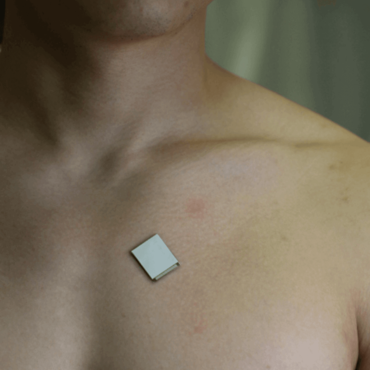
The wearable ultrasound sensor is roughly the size of a postage stamp, can be worn for up to 24 hours, and works even during strenuous exercise. (CREDIT: Xu Laboratory, UC San Diego Jacobs School of Engineering)
SAN DIEGO — A new wearable heart monitor can provide doctors with images of the organ as people go about their daily routine. The postage stamp-sized device can stay on the wearer’s chest for up to 24 hours and works even during strenuous exercise.
Researchers from the University of California-San Diego say the device uses ultrasound technology to assess both the structure and function of the heart. Cardiovascular disease is the world’s number one killer, claiming around 18 million lives annually. The sensor could prevent heart attacks or strokes and identify conditions earlier when drugs and lifestyle changes are most likely to work. It will make ultrasound more accessible to a larger population of patients.
“The technology enables anybody to use ultrasound imaging on the go,” says project leader Professor Sheng Xu in a media release.
Traditional heart monitors are too big and bulky
Currently, echocardiograms — ultrasound examinations for the heart — require highly trained technicians and bulky machines. An AI (artificial intelligence) algorithm helps the new patch to measure how much blood the heart is pumping. Most cardiovascular diseases develop because not enough blood is pumping through the organ. Issues often manifest only when the body is in motion.
Heart scans assess long-term health, detect problems as they arise, and care for critically ill patients. The new wearable, non-invasive monitor, described in the journal Nature, provides real-time, automated insights on the difficult-to-capture pumping activity — even when a person is exercising.
It uses ultrasound to continuously capture images of the four chambers of the heart in different angles and analyzes a clinically relevant subset using the custom-built AI.
“The increasing risk of heart diseases calls for more advanced and inclusive monitoring procedures,” Xu says. “By providing patients and doctors with more thorough details, continuous and real-time cardiac image monitoring is poised to fundamentally optimize and reshape the paradigm of cardiac diagnoses.”
The device also provides much better images with higher resolution and contrast than existing non-invasive methods.
“It also minimizes patient discomfort and overcomes some limitations of noninvasive technologies such as CT and PET, which could expose patients to radiation,” says Hao Huang, a PhD student in the Xu group at UC San Diego.
The patch is ideal for bodies in motion
“The device can be attached to the chest with minimal constraint to the subjects’ movement, even providing a continuous recording of cardiac activities before, during and after exercise,” explains postdoctoral researcher Xiaoxiang Gao.
Cardiac diseases are the leading cause of death among the elderly and are also becoming more prevalent among young people due to unhealthy diets and lack of exercise. The signs of cardiac diseases are transient and unpredictable, making them hard to spot. This has upped demand for more advanced, non-invasive, and cost-effective monitoring technologies such as long-term cardiac imaging.
“The heart undergoes all kinds of different pathologies,” adds postdoctoral researcher Hongjie Hu. “Cardiac imaging will disclose the true story underneath. Whether it be that a strong but normal contraction of heart chambers leads to the fluctuation of volumes, or that a cardiac morphological problem has occurred as an emergency, real-time image monitoring on the heart tells the whole picture in vivid detail and visual effect.”
The system gathers information through a wearable patch as soft as human skin, designed for optimal adherence. It sends and receives the ultrasound waves which generate a constant stream of images of the structure of the heart.
“A deep learning model automatically segments the shape of the left ventricle from the continuous image recording, extracting its volume frame-by-frame and yielding waveforms to measure stroke volume, cardiac output and ejection fraction,” concludes Mohan Li, a master’s student at UC San Diego.
Currently, the patch is connected through cables to a computer, which downloads the data automatically. However, the team has already developed a wireless circuit, which will be more appropriate for clinical use. Prof. Xu plans to commercialize the product through Softsonics, a university company spin-off.
South West News Service writer Mark Waghorn contributed to this report.


What else can they transmit and to whom?