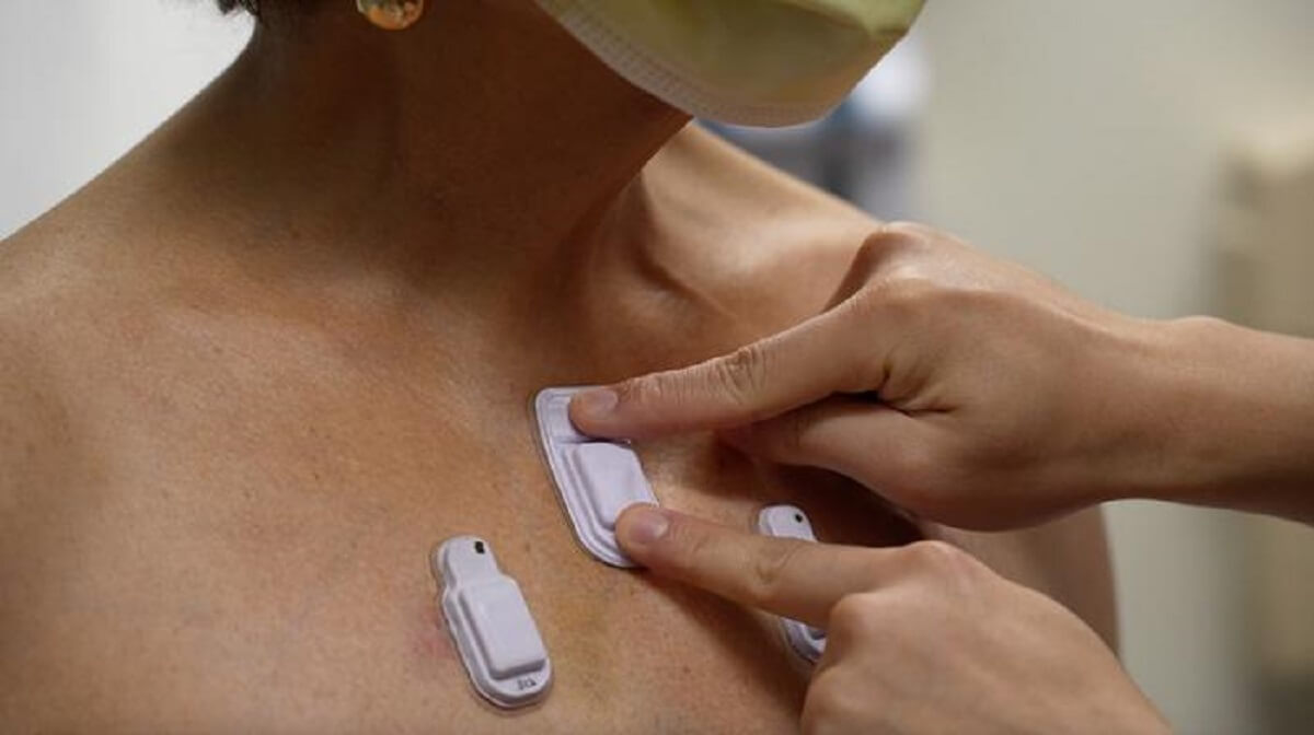
A health care worker places the wearable devices across a patient's chest to capture sounds throughout the lungs that are associated with breathing. (Credit: Northwestern University)
EVANSTON, Ill. — A revolutionary wireless acoustic device is capable of monitoring the human body with a precision that could potentially save countless lives, scientists say. Researchers crafted the wearable technology to monitor vital human organs more closely. The device, which attaches to the body, is designed to filter out ambient noise and zero in on internal sounds.
This innovation could transform health monitoring, particularly for premature infants and adults with respiratory diseases.
The medical experts behind the device acknowledge that it outperforms the traditional methods used by multiple doctors in patient health assessment. Doctors routinely listen to the sounds of the body — breaths, heartbeats, and the movement of food through the gastrointestinal tract — as indicators of health. However, these sounds can be subtle, hard to track, and easily overwhelmed by background noise.
Researchers at Northwestern University developed miniature wearable devices that surpass the intermittent insights gained from standard medical examinations. These devices adhere softly to the skin, providing continuous monitoring and wireless tracking of sounds across various regions of the body.

In initial studies, the devices were tested on 15 premature infants with respiratory and intestinal issues and 55 adults, including many with chronic lung diseases. The devices not only met clinical-grade accuracy standards but also introduced new functions not yet seen in research or clinical practice.
“Physicians have to put a conventional, or a digital, stethoscope on different parts of the chest and back to listen to the lungs in a point-by-point fashion,” says John Rogers, a bioelectronics pioneer at Northwestern University who spearheaded the device’s development, in a media release.
The researchers liken the device to having 13 doctors listening to the patient’s body simultaneously.
“The idea behind these devices is to provide highly accurate, continuous evaluation of patient health and then make clinical decisions in the clinics or when patients are admitted to the hospital or attached to ventilators,” says Dr. Ankit Bharat, a thoracic surgeon at Northwestern Medicine who conducted the clinical research on adult subjects. “A key advantage of this device is to be able to simultaneously listen and compare different regions of the lungs.”
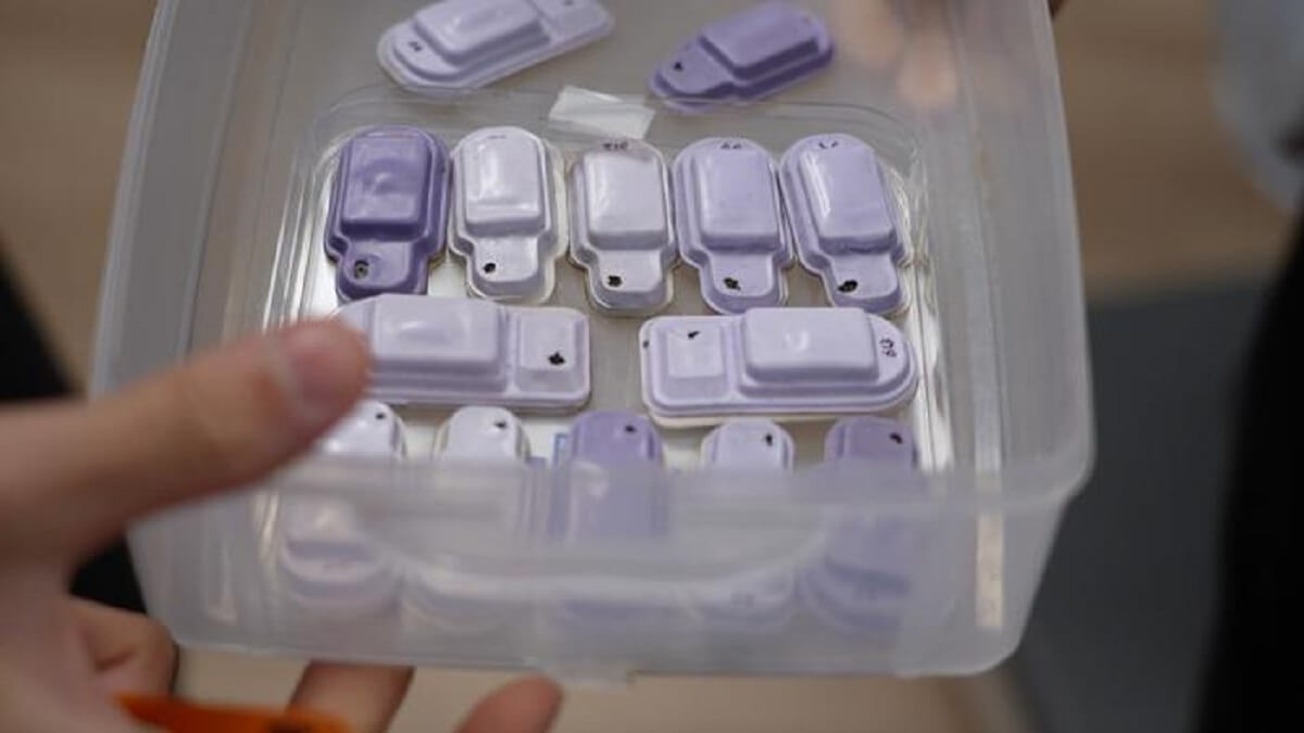
The compact devices are equipped with high-performance digital microphones and accelerometers and adhere gently to the skin, forming a comprehensive sensing network. They capture sounds and correlate them with bodily processes, mapping the flow of air in the lungs, cardiac rhythm changes, and the movement of food and gas through the intestines.
Each device, encased in soft silicone, is approximately 40 millimeters in length, 20 millimeters wide, and eight millimeters thick. Despite its small size, it contains flash memory, a battery, Bluetooth capabilities, and two microphones — one facing the body and the other outward. An algorithm differentiates between internal and external sounds.
“Lungs don’t produce enough sound for a normal person to hear. They just aren’t loud enough, and hospitals can be noisy places,” Dr. Bharat explains. “When there are people talking nearby or machines beeping, it can be incredibly difficult. An important aspect of our technology is that it can correct for those ambient sounds.”
Capturing ambient sounds also provides context about the patient’s environment, which is crucial for treating premature infants.
“It also offers immediate opportunities to address any sources of stressful or potentially compromising auditory stimuli,” notes co-first author Dr. Wissam Shalish, a neonatologist at Montreal Children’s Hospital.
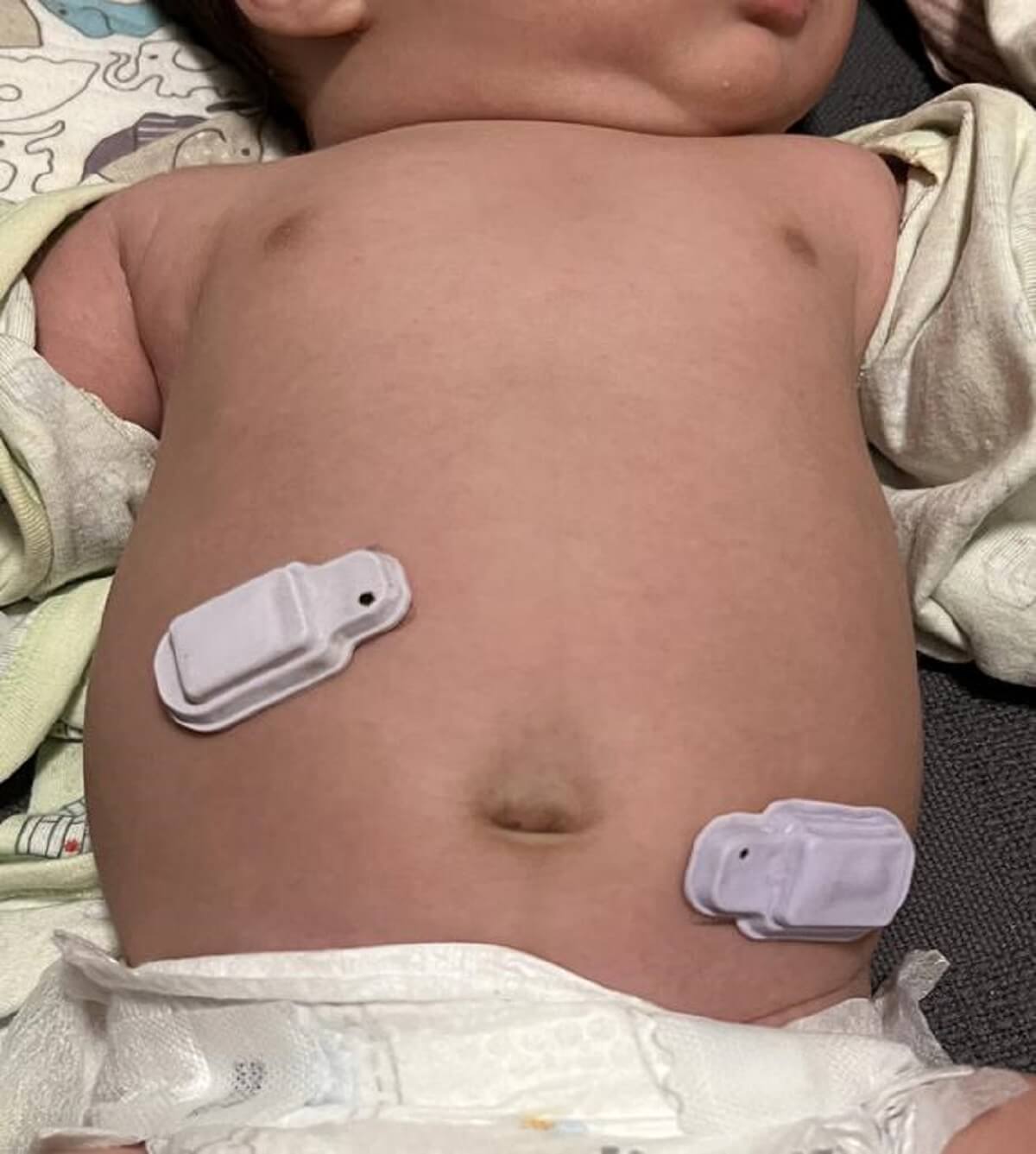
The scientists developed the devices with two at-risk patient groups in mind: premature infants in NICUs and post-surgery adults. Premature babies, particularly those born before the respiratory system matures at around 28 weeks, often face breathing complications, such as apnea, leading to extended hospital stays or even death.
“Many of these babies are smaller than a stethoscope, so they are already technically challenging to monitor,” co-author Dr. Debra Weese-Mayer points out.
In both children and adults, cardiorespiratory and gastrointestinal issues are leading causes of death within the first five years of life. Gastrointestinal problems, indicated by diminished bowel sounds, can signal digestion issues and potential obstructions.
Sensors were placed on premature infants across their abdomens, with early results matching adult intestinal motility measurements, the current standard of care.
The devices captured lung sounds and body movements at various locations, allowing for the analysis of individual breaths across the lungs.
“With these wireless sensors, we can capture different regions of the lungs and assess their specific performance and each region’s performance relative to one another. “Eventually we can personalize treatments to individual patients,” says Dr. Bharat.
The study is published in the journal Nature Medicine.
You might also be interested in:
- Wireless wearable sensor that monitors organ health has ‘potential to save and improve lives’
- Best Fitness Trackers For 2024: Top 5 Wearable Tech Devices Recommended By Experts
- Motion sensors placed throughout home can detect heart problems, cognitive decline
South West News Service writer James Gamble contributed to this report.


