LEXINGTON, Ky. — A 60-year-old mystery surrounding the heart has finally been cracked. University of Kentucky College of Medicine scientists were able to map out a crucial part of the heart’s molecular composition to help better understand the organ’s intricate structure.
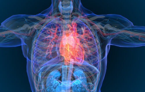
In this study, researchers explored the microscopic intricacies of the heart, examining a component known as myosin filaments, which play a fundamental role in heart muscle function.
The human heart consists of billions of cells, each containing thousands of smaller structures known as sarcomeres. These sarcomeres are essentially the building blocks of muscle within the heart. Within each sarcomere, there are hundreds of myosin filaments. To visualize the scale of their work, scientists liken it to examining single strands of hair on a continent-sized heart.
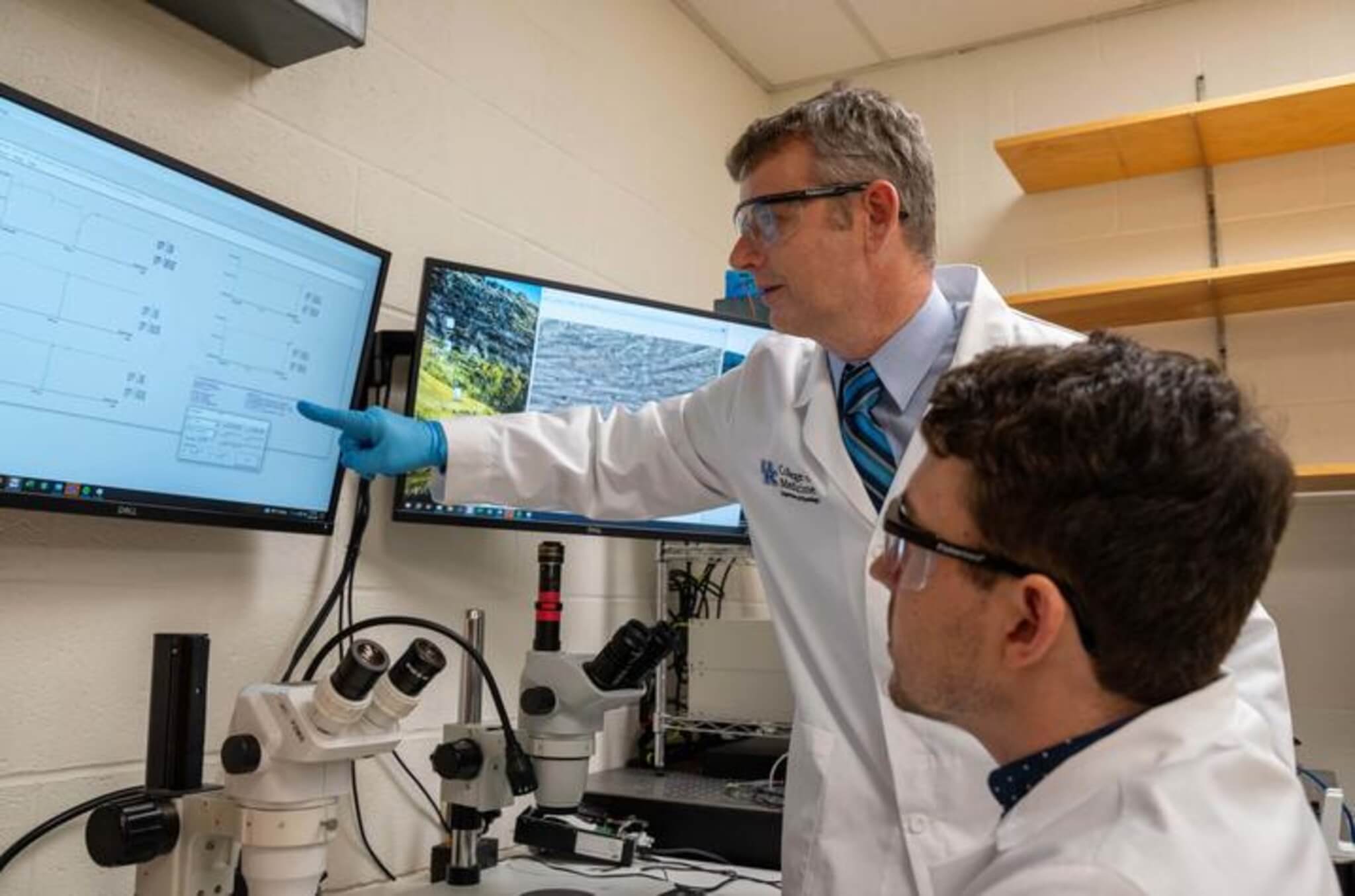
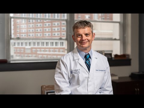
“Each filament has roughly 2,000 molecules arranged in a really complicated structure that scientists have been trying to understand for decades,” says study author Dr. Kenneth Campbell, the director of translational research in the Division of Cardiovascular Medicine in the UK College of Medicine, in a university release. “We knew quite a lot about the individual molecules and people thought the myosins could be arranged in groups of six that were called crowns, but not much beyond that.”
What sets their discovery apart is the revelation that there are three distinct types of these “crowns” within the myosin filaments, each with unique interactions. This finding suggests that the heart muscle’s control is more precise than previously believed. Additionally, they observed the arrangement of myosin-binding protein-C, another protein linked to genetic heart disease, within this complex structure. This newfound insight provides a deeper understanding of how molecules are organized within the heart.
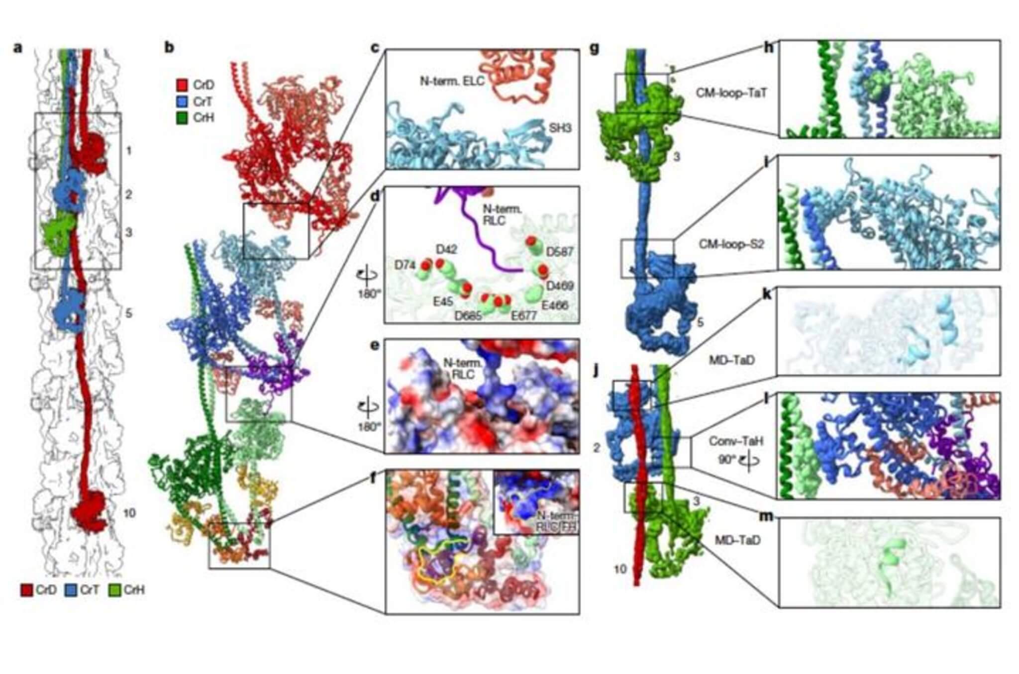
To accomplish this groundbreaking work, Dr. Campbell collaborated with researchers at the University of Massachusetts Chan Medical School. Together, they produced detailed 3D reconstructions of cardiac thick filaments, offering a fresh perspective for interpreting structural, physiological, and clinical observations.
The significance of this research extends beyond the laboratory.
“This study is important for discovering new drug therapies for heart disease which Kentucky desperately needs,” notes Dr. Campbell. “It gives us a much better understanding of how the molecules in our hearts interact.”
Heart disease is a pressing concern in Kentucky, where it is the leading cause of death, placing the state among the top 10 in the nation for the highest death rate due to this disease, according to the Centers for Disease Control and Prevention (CDC).
“We’re interested in therapies for different kinds of heart failure and myopathies, where the heart muscles don’t work very well,” explains Dr. Campbell. “Our research is one of many projects underway at the university to help come up with better therapies for heart disease.”
The heart samples used for this research were collected from the Gill Cardiovascular Biorepository, of which Dr. Campbell is the director. These samples are generously donated for research purposes by patients who receive cardiovascular care at the University of Kentucky.
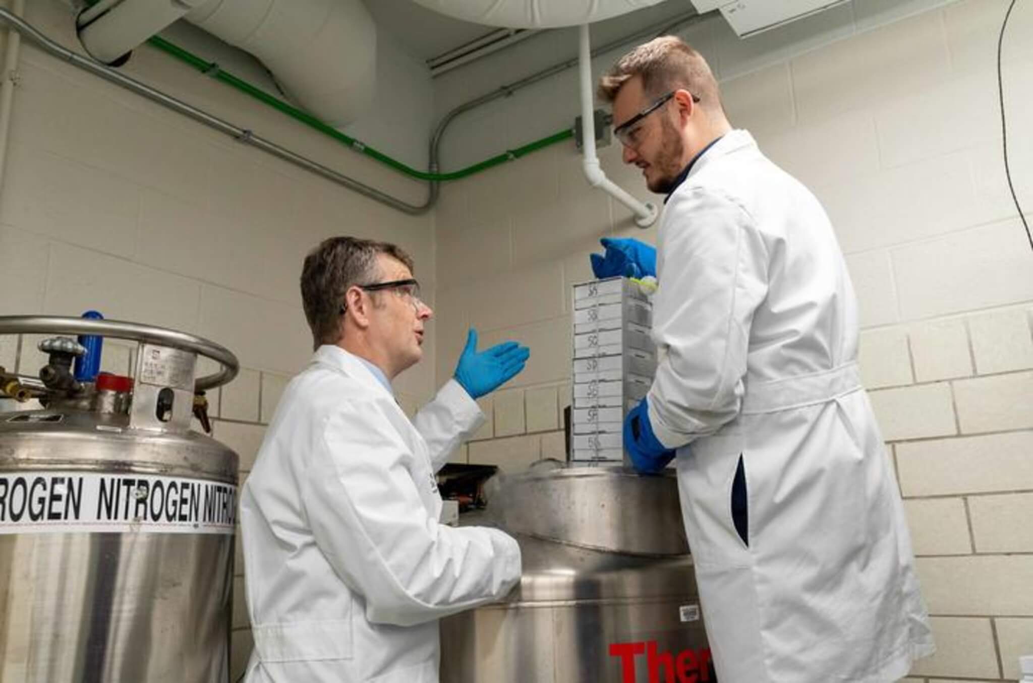
“We started the Gill Cardiovascular Biorepository in 2008. With the help of a surgeon at UK HealthCare, we started collecting samples of myocardium from organ donors and from patients who were getting cardiac transplants,” says Dr. Campbell. “Now we’ve built a huge resource with roughly 15,000 samples from nearly 500 people.
Dr. Campbell, who holds a joint appointment in cardiovascular medicine and physiology, has transformed his background in physics into a career focused on making a tangible difference in people’s lives.
“I used to care a lot about math and molecules,” says Dr. Campbell. “But after hearing a friend who’s a cardiothoracic surgeon talk about patients, I realized I could take my scientific skills and do research that has a chance of helping people. It’s given my science purpose.
The study is published in the journal Nature.
You might also be interested in:
- Fainting mystery solved! Genetic discovery connects heart and brain when people pass out
- Don’t snooze on this! Study reveals mild sleep deprivation could cause heart disease
- Best Ways To Improve Heart Health: Top 5 Natural Methods To Care For Your Ticker, Per Health Experts