
(© natali_mis - stock.adobe.com)
LONDON — Around the world, people die from heart conditions they didn’t even know they had. Researchers from the University College London have studied one called hypertrophic cardiopathy (HCM). Their work shows that combining two heart scans could help doctors detect the deadly condition before symptoms arise and signs on conventional tests emerge.
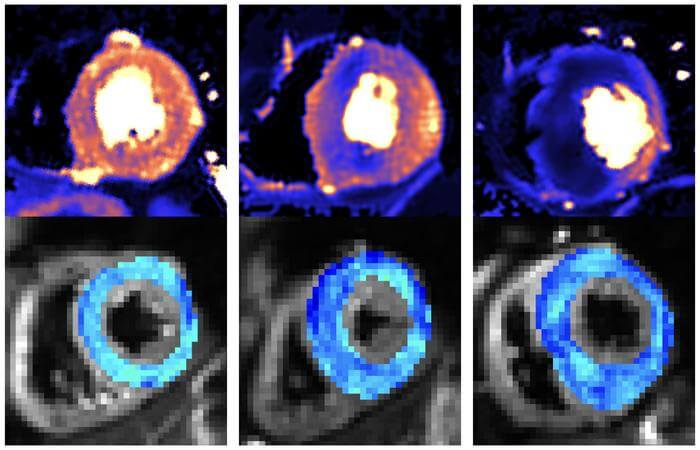
HCM is a deadly condition affecting the left ventricle of the heart, which is the one that pumps blood to the rest of the body. The team split the participants into three groups: healthy people, people who already had HCM, and people with an HCM-causing genetic mutation but no overt signs of disease, like muscle heart thickening. The two tests they used are cardiac diffusion tensor imaging (cDTI) — which is a type of MRI scan that shows how individual heart muscle cells are structured and packed together — and cardiac MRI perfusion (perfusion CMR), which detects problems with the small blood vessels that supply the heart muscle.
The scans can successfully show that people with overt signs of HCM have abnormal heart muscle cell organization, as well as a high rate and severity of microvascular disease compared to healthy participants. Even more importantly, the scans could detect abnormalities in the structure of people with a problematic gene who failed to display symptoms. They discovered that 28 percent of them had defects in their blood supply in comparison to the healthy group. Doctors can therefore more accurately detect early signs of HCM.
“By linking advanced imaging to our cohort of HCM patients (and relatives) with extensive genetic testing, this study detected microstructural abnormalities in vivo in mutation carriers for the first time and was the first to compare these parameters in HCM patients with and without a causal mutation,” says Dr. Luis Lopes (UCL Institute of Cardiovascular Science), senior author of the study, in a university release.
Mavacamten, the first drug to slow HCM progression, has been recently approved in Europe. This will allow doctors to lessen disease severity once symptoms start to appear. There are also genetic therapies in development that help prevent symptoms as a whole, by halting HCM development early on.
“The ability to detect early signs of HCM could be crucial in trials testing treatments aimed at preventing early disease from progressing or correcting genetic mutations. The scans could also enable treatment to start earlier than we previously thought possible,” says Dr. George Joy, who led the research with Professor James Moon and Dr. Luis Lopes.
“We now want to see if we can use the scans to identify which patients without symptoms or heart muscle thickening are most at risk of developing severe HCM and its life-changing complications. The information provided from scans could therefore help doctors make better decisions on how best to care for each patient.”
The findings are published in the journal Circulation.


